Introduction
Maxillary cancer is a malignant tumor affecting the maxillary bone, characterized by infiltrative growth and regional metastasis. The main symptoms of neoplasia are nasal congestion, purulent bloody discharge, constant pain of a nagging nature, pathological mobility of intact teeth, deformation of the alveolar process. The diagnosis of maxillary cancer is made based on the patient's complaints, physical examination data, the results of radiography, cytologic and pathohistological studies. Treatment of maxillary cancer is combined; resection of the maxillary bone is preceded by a course of chemotherapy or radiation therapy.
General information
Maxillary cancer is a primary or metastatic malignant neoplasm affecting the upper jaw. Among patients presenting to an oncology clinic, upper jaw cancer is diagnosed three times more often than mandibular bone cancer. There are four cases of cancer of the same localization per one case of sarcoma of the upper jaw. The main group of patients is middle-aged people (40 to 60 years old). In 65% of cases, cancer of the upper jaw is detected after the age of 50. Most often the tumor develops from the mucous membrane of the maxillary sinus. Histogenetically malignant neoplasm of the upper jaw in 80% of cancer patients is a squamous cell cancer. Metastasis of maxillary cancer is observed late, metastasis is detected in every third patient.
Causes of maxillary cancer
The most common causes of maxillary cancer are chronic sluggish inflammatory diseases of the maxillary sinuses. Less often, a malignant tumor develops directly in the bone tissue from the epithelial islets of Malasse, or grows into the bone from the oral mucosa covering the palate, cheeks, alveolar outgrowth. High risks of malignization in case of chronic trauma to the mucosa with sharp edges of destroyed teeth, unpolished base of removable dentures or protruding ribbed components of orthopedic or orthodontic appliances.
In addition to the primary malignant neoplasm, secondary forms have also been described in dentistry, when maxillary cancer developed as a result of metastasis of tumors of the mammary gland, thyroid gland, stomach in cancer patients. Metaplastic changes in the cylindrical epithelium lining the maxillary sinus occur as a result of chronic maxillary sinusitis. The formation of hyperplastic overgrowths is accompanied by tissue malignization. Histologically, cancer of the upper jaw in the predominant majority of cases is squamous cell keratinizing.
Classification of upper jaw cancer
The following stages of cancer of the upper jaw are distinguished:
- Stage 1. The tumor is localized in one anatomical region. Destructive changes in the bone are absent.
- Stage 2a. Cancer of the upper jaw does not spread to neighboring areas. Within the anatomical region of the location of the neoplasm, destructive changes of the bone are present.
- Stage 2b. Cancer of the upper jaw sprouts into adjacent areas. On the side of the lesion, a single unconnected metastasis with surrounding tissues is detected.
- Stage 3a. Cancer of the upper jaw spreads to the orbit, nasal cavity. Signs of lesions are detected on the palate, alveolar process. There is osteolysis of bone tissue.
- Stage 3b. In the course of examination, unilateral or bilateral metastases are detected.
- Stage 4a. Cancer of the upper jaw spreads to the nasopharynx, the base of the skull. There are ulcerations on the skin. There are no distant metastases.
- Stage 4b. Cancer of the upper jaw affects adjacent areas. Metastases fused with surrounding tissues are also found.
Symptoms of maxillary cancer
In the initial stages, maxillary cancer is asymptomatic. During the first 2-3 months, patients associate nasal congestion, scanty serous discharge with manifestations of chronic maxillary sinusitis. Further, the clinic of maxillary cancer becomes more obvious. Symptomatology directly depends on the localization of the pathological focus. If the malignant neoplasm develops in the upper part of the medial wall, the lacrimal sac and lacrimal passages are involved in the pathological process, which further leads to the appearance of signs of secondary dacryocystitis. Patients have increased lacrimation, corresponding to the side of the lesion. The skin in the area of the inner corner of the eye becomes edematous, hyperemic.
With the progression of cancer of the upper jaw, the lower ocular wall is destroyed, as a result of which exophthalmos and diplopia develop, visual acuity decreases. If the neoplasm is localized in the lower part of the medial wall, patients indicate the presence of brown discharge from the nose with an unpleasant putrid odor. There is a feeling of heaviness, nasal congestion. If maxillary cancer affects the anterior wall of the maxillary sinus, there is a pronounced pain syndrome in the area of intact premolars or molars. Further, pathologic mobility of the teeth joins.
Severe neuralgic pain is also observed with the localization of maxillary cancer in the upper-outer quadrant. When the tumor process spreads to the area of masticatory muscles, mouth opening is disturbed. As a result of compression of the venous plexus, the outflow of lymph from the retrobulbar tissue is blocked, which further leads to chemosis and exophthalmos. An abbreviated clinical picture is observed when maxillary cancer develops in the lower part of the external wall of the maxillary sinus. The main complaints are similar to the manifestations of chronic maxillary sinusitis.
Diagnosis of maxillary cancer
The diagnosis of cancer of the upper jaw is made on the basis of patient complaints, anamnesis data, the results of clinical, radiologic, cytologic and pathohistological studies. At the initial stages, the clinic of maxillary cancer is similar to chronic maxillary sinusitis, ethmoiditis. If the tumor process spreads to the bone tissue from the mucous membrane of the palate, cheek or alveolar outgrowth, during examination, the dentist identifies the initial pathological focus in the oral cavity.
With the exophytic type of growth, the tumor is represented by overgrowths with signs of ulceration. Crater-shaped ulcerated surface is found with endophytic growth of malignant neoplasm. At late stages of maxillary cancer, deformation of the alveolar process, pathological mobility of intact teeth, absence of pain and tactile sensitivity in the area of tumor localization are detected. Sprouting of the malignant neoplasm into the orbit leads to the development of exophthalmos and causes loss of vision.
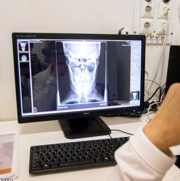
Radiographically, in the initial stages of cancer of the upper jaw, developing from the mucous membrane of the maxillary sinus, veiling of the sinus is revealed. In the area of the suture connecting the alveolar process to the zygomatic bone, as well as in the area of the lower glabellar cleft, destructive changes of the bone, uncharacteristic of chronic maxillary sinusitis, are detected.
To confirm the diagnosis of cancer of the upper jaw, laboratory tests are used. The presence of a malignant tumor is signaled by the detection in the course of cytological analysis of tumor cells in the lavage waters obtained from the maxillary sinuses. In the absence of drainage through the maxillary sinus junction, puncture is performed. The obtained material is sent for pathohistological examination. Upper jaw cancer is differentiated with chronic maxillary sinusitis, odontogenic radicular cyst, chronic osteomyelitis. Physical examination is performed by a dental surgeon, an oncologist, and if necessary, an otolaryngologist.
Maxillary cancer treatment
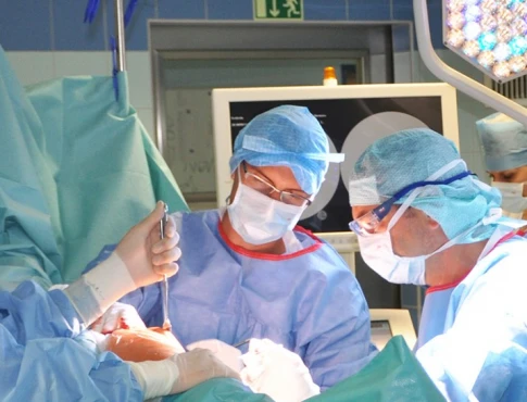
If the cancer of the upper jaw has not spread to the base of the skull, treatment includes gamma therapy or chemotherapy accompanied by the injection of drugs into the regional arteries. Extirpation of the maxilla is performed after 3 weeks. After surgery, contact radiation therapy of the bed of the removed neoplasm is indicated. If metastasis of maxillary cancer is suspected, excision of cervical lymph nodes together with fiber is performed.
The presence of several nonadherent metastases with surrounding tissues or one adherent metastasis is a direct indication for radical intervention consisting in removal of lymph nodes together with subcutaneous tissue, cervical section of the internal jugular vein, mandibular salivary gland and sternoclavicular-papillary muscle. The surgical defect is filled with a plate removable prosthesis (provided that soft tissues are preserved) or an exoprosthesis is made. Closure of the mouth cavity with the nasal cavity is carried out at the expense of the obturator plate. If there are signs of involvement of the skull base in the pathological process, surgical intervention is not performed. The basis of treatment of maxillary cancer in this case is chemotherapy and radiation therapy.
With early detection of malignant neoplasm, the prognosis is favorable. In the late stages of maxillary cancer, the spread of tumor cells to the base of the skull is accompanied by metastasis. In this case, the prognosis for life is unfavorable. After radical lymphadenectomy, venous outflow worsens, there is a persistent deformity of the neck, which negatively affects the quality of life of patients.
