Overview
Esophageal cancer (EC) is the seventh most commonly diagnosed malignancy worldwide and the sixth most common cause of cancer-related death. [1] Due to the aggressive nature of EC, the 5-year overall survival rate is 30–40%. [2] However, esophageal cancer is quite treatable.
Where does esophageal cancer start?
The esophagus is a part of the digestive system. It is the 25 cm long fibromuscular tube that passes food and liquid from the throat to the stomach. [3]
The esophageal wall is a four-layer structure with a mucosa, submucosa, muscularis propria, and adventitia. [4]
The origin of malignant tumors comes from the cells of the inner layer - the mucosa, which consists of 2 main types of cells:
- the glandular cells that produce mucus. They give rise to adenocarcinoma.
- the squamous epithelial cells lining the esophagus. They give rise to squamous cell carcinoma.[4] [5]
Adenocarcinoma and squamous cell carcinoma occur more often. A third, rarer type of esophageal cancer is small cell carcinoma. It begins in neuroendocrine cells, which release hormones into the bloodstream in response to nerve signals.
Other types of esophageal cancer, such as small-cell neuroendocrine cancers, lymphomas, and sarcoma, are rare and comprise less than 1% of esophageal cancers.[5] [6]
Risk factors
A risk factor is anything that increases a person’s chance of developing cancer. Although risk factors often influence the development of cancer, most do not directly cause cancer. Some people with several risk factors never develop cancer, while others with no known risk factors do. The following factors may raise the risk of developing esophageal cancer: [5]
- Age between 45 and 70;
- Gender. Men are 3 to 4 times more likely to develop esophageal cancer than women;
- Race. Black people are twice as likely as white people to develop the squamous cell type of esophageal cancer;
- Tobacco. Using any form of tobacco, such as cigarettes, cigars, pipes, chewing tobacco, and snuff raises the risk of esophageal cancer;
- Alcohol. Heavy drinking over a long period increases the risk of squamous cell carcinoma of the esophagus, especially when combined with tobacco use;
- Gastroesophageal reflux disease (GERD) is a condition in which stomach acid repeatedly flows back up into the esophagus;
- Barrett’s esophagus is a condition in which the flat pink lining of the esophagus becomes damaged by acid reflux, which causes the lining to thicken and become red. Damage to the lining of the esophagus causes the squamous cells in the esophagus lining to turn into glandular tissue. People with Barrett's esophagus are more likely to develop adenocarcinoma of the esophagus.
- A diet with low fiber;
- Obesity. Patients with obesity have an increased risk of GERD. Also, adipose tissue cells secrete inflammatory factors that adversely affect the condition of the mucous layer of the esophagus;
- Lye. Accidental ingestion of the lye that can be found in some cleaning products, such as drain cleaners;
- Achalasia. Achalasia is a condition when the lower muscular ring of the esophagus does not relax during the swallowing.
Symptoms
Esophageal cancer may not cause symptoms early on; it usually happens when the disease is advanced. These symptoms of esophageal cancer are associated with tumor growth, mechanical obstruction in the esophagus, or destruction of surrounding tissues and organs. [4] [5]
Symptoms of esophageal cancer include:
- swallowing problems (dysphagia)
- pain in your throat or the middle of your chest, especially when swallowing
- choking, nausea, and vomiting
- heartburn or acid reflux
- symptoms of indigestion, such as excessive burping
- a cough that is not getting better
- a hoarse voice
- bad breath
- black stool or coughing up blood (although these are uncommon)
- loss of appetite or unexplained weight loss
- feeling tired or having no energy
- feeling sick
Diagnosis
The primary techniques used to diagnose suspected esophageal cancer include:[6] [7]
- A barium swallow or contrast esophagram is an imaging test that examines the upper gastrointestinal tract, specifically the esophagus, throat, and back of the mouth. A barium swallow can help diagnose structural or functional issues. Barium is a dry powder that is mixed with water to create a drinkable contrast material that must be taken orally, after which a series of X-rays are taken;
- Esophagogastroscopy. The doctor passes an endoscope, a thin, lighted tube, into the esophagus to examine it. Endoscopic ultrasound uses sound waves to provide more information about the extent of tumor involvement in nearby tissues.
- Biopsy: during the endoscopy, the doctor can take tissue samples from suspicious esophagus areas, followed by histological examination under a microscope. Doctors also examine the tumor sample for the presence of proteins that can be affected by targeted drugs.
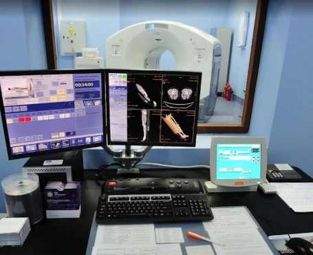
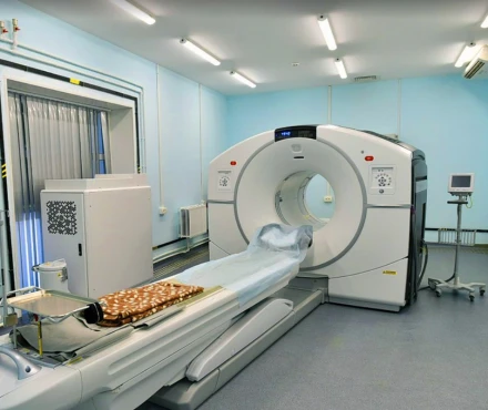
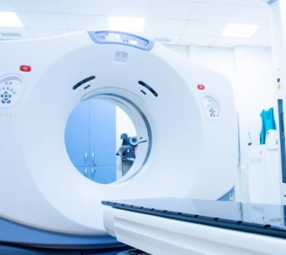
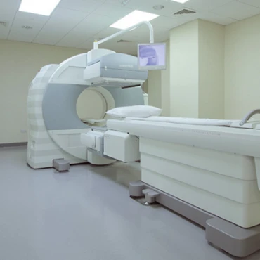
Other tests, including computed tomography (CT) scans, positron emission tomography (PET) scan, thoracoscopy, and laparoscopy, may be performed to determine if the cancer has spread, or metastasized, outside of the esophagus. This process is called "staging" and is necessary for planning treatment.
Staging
There are different ways of staging esophageal cancer. TNM and number staging systems are most commonly used in clinical practice. [4]
TNM staging system evaluates:
T - the size of the primary tumor
N - involvement of lymph nodes in the tumor
M - presence of metastases.
The number staging system includes five stages, which are:
0 - high-grade dysplasia: abnormal cells (not yet cancer) are found only in the layer of cells that line the esophagus;
I - cancer cells are found only in the layer of cells that line the esophagus.
II - the cancer has reached the muscle layer or the outer wall of the esophagus. In addition, the cancer may have spread to 1 to 2 nearby lymph nodes.
III - the cancer has reached deeper into the inner muscle layer or the connective tissue wall. It may have spread beyond the esophagus into surrounding organs and/or has spread to more local lymph nodes.
IV - the most advanced stage, where cancer has spread to distant organs and/or lymph nodes far from the esophagus.
Treatment
Esophageal cancer is quite a treatable type of cancer. Treatment for esophageal cancer typically includes a combination of surgery, radiation therapy, and chemotherapy.
The choice of treatment regimen depends on several factors, such as:
histological type of cancer
size and tumor location
presence of metastases
patient's health
Surgery
Surgery is the most effective treatment for early-stage esophageal cancer.
If the tumor hasn't spread deeply into the tissue, ablation techniques can be used. These include laser therapy, photodynamic therapy (PDT), radiofrequency ablation, argon plasma ablation (APC), and cryoablation. These methods can be combined with endoscopic resection.
Endoscopic mucosal resection (EMR) and endoscopic submucosal dissection (ESD) are good options for lesions that do not penetrate the mucosal layer (T1a) or the lesions that develop only within the epithelium and lamina propria. Endoscopic methods are safe and have a minimal number of complications. These methods are characterized by a short recovery time. [3]
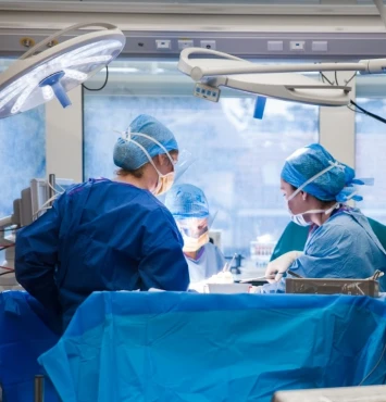
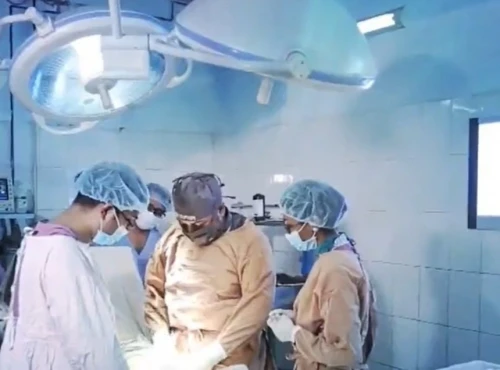
An esophagectomy is the most effective treatment for patients without invasion to adjacent organs or distant metastasis. It involves removing some or most of the esophagus. Depending on the tumor's location and clinic equipment, an esophagectomy can be open or minimally invasive. [4] During an open esophagectomy, the surgeon removes all or part of the esophagus through an incision in the neck, chest, abdomen, or a combination of these areas. The esophagus is then reconstructed using another organ, such as the stomach or the small or large intestine. Minimally invasive esophagectomy includes laparoscopy or robot-assisted techniques. Sometimes, a combination of these approaches may be used. When possible, these procedures are performed through several small incisions, leading to less pain and quicker recovery compared to traditional surgery. [4] [5]
An esophagogastrectomy is used when cancer occurs in the middle or lower part of the esophagus, or it has grown into the stomach. In such cases, removing the top part of the stomach and a part of the esophagus is necessary. An esophagogastrectomy is always accompanied by the need to reconstruct the removed part of the esophagus using part of the intestine. [5]
Lymphadenectomy or lymph node removal is often done during an esophagectomy. Removing the lymph nodes from around the esophagus and examining them for tumor metastases is important. The number of lymph nodes the surgeon removes varies. [4]
The main postoperative issue is restoring nutrition. A gastrostomy tube, surgically placed through the abdominal wall into the stomach, helps the patient continue feeding. This tube can remain in place until the end of therapy.[6]
Radiotherapy
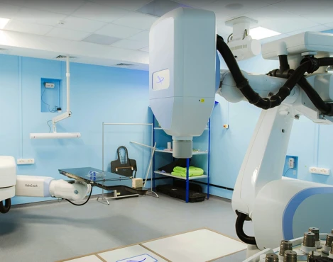
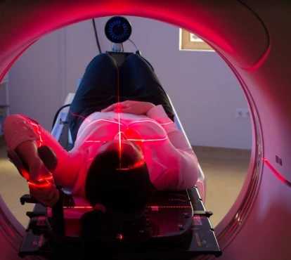
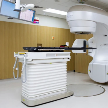
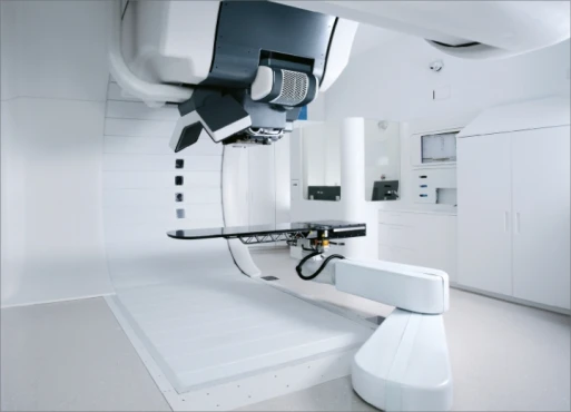
Two types of radiation therapy are used for esophageal cancer: external beam radiation therapy (EBRT) and brachytherapy. Brachytherapy is a type of internal radiation therapy in which a source of radiation is placed in or near the tumor. External beam radiation therapy is used in combination with chemotherapy. This option reduces the radiation dose and intensity of its adverse effects.
Radiotherapy alone is not standard for esophageal cancer due to its high toxicity and limited effectiveness. However, it can be used to control symptoms in advanced esophageal cancer that has spread or in combination with chemotherapy (chemoradiotherapy).
Radiation therapy can be carried out before surgery to reduce tumor size and surgery volume. In advanced stages, when cancer is unresectable, chemoradiotherapy on its own can be the primary treatment. [7]
Systemic therapy
Systemic therapy includes chemotherapy, targeted therapy, and immunotherapy.
Chemotherapy kills fast-growing cells throughout the body, including cancer cells and some normal cells. When chemotherapies are combined, it is called multi-agent or combination chemotherapy. Chemotherapy might be given before surgery (neoadjuvant chemotherapy) and/or after surgery (adjuvant chemotherapy). Long-term multicenter trials confirm the effectiveness of neoadjuvant chemoradiation for patients with squamous cell carcinoma.
Targeted therapy focuses on specific or unique features of cancer cells. It examines how cancer cells grow, divide, and move in the body. These drugs stop or inhibit the action of molecules that help cancer cells grow and/or survive. They are often selected based on next-generation sequencing (NGS) and immunohistochemistry (IHC) results, which are performed on tumor biopsy samples. Several targeted therapies have shown promise in pairing with radiation in preclinical and clinical studies, including epidermal growth factor receptor (EGFR) inhibitors, receptor tyrosine kinase (RTK) inhibitors, cell cycle checkpoint inhibitors (Wee1, Chk1/2), and vascular endothelial growth factor (VEGF) inhibitors, among others.
Immunotherapy can be given alone or with other types of treatment. Immunotherapy is a systemic treatment that tries to reactivate your own immune system against cancer. Immunotherapy may also improve outcomes in the neoadjuvant setting when combined with chemoradiation.[11]
Complications of Treatment
Despite its high effectiveness, the treatment of esophageal cancer is very aggressive and can lead to various complications.
The most common are:
- Functional gastric emptying disorder is defined as delayed gastric emptying in the absence of mechanical obstruction of the stomach;
- Reflux esophagitis is associated with the backflow of acid from the stomach into the esophagus.
- Pulmonary complications
- Chylothorax is a condition where lymphatic fluid, called chyle, accumulates in the pleural cavity, the space between the lungs and the chest wall. It is more often caused by damage to the thoracic duct during surgery.
- Anastomotic or thoracogastric fistula develops due to failure of the suture at the site of resection of the esophagus;
- Anastomotic stenosis or esophageal stenosis, in most cases, is caused by poor healing of the mucous membrane;
- Severe diarrhea: esophageal cancer treatment may also lead to severe diarrhea by causing gastrointestinal dysfunction;
- Radiation damage to the mucous membrane of the oral cavity and esophagus leads to ulceration and severe pain.
Prognosis
Survival depends on several factors, primarily the stage of the disease. Patients with very early-stage disease have a better survival chance. Among those who receive treatment, 40% will survive one year or more, 15% will survive up to five years, and 10% will survive ten years or more. [9]
People with a low body mass index and severe nutritional deficiencies are at risk for developing severe complications of therapy and its interruption. [9]
References
1. Yang J, Liu X, Cao S, Dong X, Rao S, Cai K. Understanding esophageal cancer: the challenges and opportunities for the next decade. Front Oncol. 2020;10:1727.
2. Ferlay J, Soerjomataram I, Dikshit R, Eser S, Mathers C, Rebelo M, et al. Cancer incidence and mortality worldwide: sources, methods and major patterns in GLOBOCAN 2012. Int J Cancer. 2015;136(5):E359–86.
3. Bajwa SA, Toro F, Kasi A. Physiology, Esophagus. [Updated 2023 May 1]. In: StatPearls [Internet]. Treasure Island (FL): StatPearls Publishing; 2024 Jan.
4. Squier CA, Kremer MJ. Biology of oral mucosa and esophagus. J Natl Cancer Inst Monogr. 2001;(29):7-15.
5. Domper Arnal MJ, Ferrández Arenas Á, Lanas Arbeloa Á. Esophageal cancer: Risk factors, screening and endoscopic treatment in Western and Eastern countries. World J Gastroenterol. 2015 Jul 14;21(26):7933-43.
6. Abbas G, Krasna M. Overview of esophageal cancer. Ann Cardiothorac Surg. 2017 Mar;6(2):131-136.
7. D'Journo XB, Thomas PA. Current management of esophageal cancer. J Thorac Dis. 2014 May;6 Suppl 2(Suppl 2):S253-64.
8. Bolger, J.C., Donohoe, C.L., Lowery, M. et al. Advances in the curative management of oesophageal cancer. Br J Cancer 126, 706–717 (2022).
9. Tustumi F, Kimura CM, Takeda FR, Uema RH, Salum RA, Ribeiro-Junior U, Cecconello I. Prognostic factors and survival analysis in esophageal carcinoma. Arq Bras Cir Dig. 2016 Jul-Sep;29(3):138-141
10. Vitzthum LK, Hui C, Pollom EL, et al. Trimodality Versus Bimodality Therapy in Patients With Locally Advanced Esophageal Carcinoma: Commentary on the American Society of Clinical Oncology Practice Guidelines. Pract Radiat Oncol 2021;11:429-33.
11. Kelly RJ, Ajani JA, Kuzdzal J, et al. Adjuvant Nivolumab in Resected Esophageal or Gastroesophageal Junction Cancer. N Engl J Med 2021;384:1191-203.


