Disease Types & Epidemiology
How common is the disease?
Kidney or renal cancer originates in the tissues of the kidneys. Most kidney cancers are lesions that start in cells lining the inner or outer surfaces of the body. In the kidneys, carcinomas most often arise from the cells lining the renal tubules.
Approximately 9 out of 10 kidney cancers are renal cell carcinoma (RCC), accounting for about 4% of all cancer cases in the United States.
In 2024, an estimated 81,800 new kidney cancer cases will be diagnosed, and around 14,890 people are expected to be diagnosed with the disease, according to SEER data [NCI, 2024]. The median age at diagnosis is 64 years, and the condition is more prevalent in men than women, with a ratio of roughly 2:1.
RCC may manifest as multiple tumors within a single kidney or involve both kidneys. As the most common type, RCC is often simply referred to as kidney cancer.
Other, less frequent cancers can develop in the kidney. While they can sometimes be mistaken for RCC, they are treated differently.
- Urothelial carcinoma originates in the cells lining the renal pelvis and ureter that drain the kidney. Although these tumors may be found near the kidneys, they are not kidney tumors themselves. These tumors share many similarities with bladder cancer and were previously called transitional cell cancer.
- Wilms tumor typically affects young children and involves low-differentiated cancer cells in the kidney's parenchyma.
- Renal sarcoma starts in the blood vessels or connective tissue of the kidney.
Renal cell carcinoma is the most common type of kidney cancer. RCC can be classified into subtypes by examining the cancer cells under a microscope. This process is called histology. Tumor histology and other factors are crucial in selecting the appropriate treatment plan.
1. Clear cell RCC is the most prevalent subtype in about 7 out of 10 RCC cases. These cancer cells appear very pale or clear under the microscope.
2. Non-clear cell RCC, known as nccRCC, encompasses several categories: 2.1. Papillary RCC is the most common nccRCC subtype. Most papillary tumors resemble long, thin, finger-like growths when viewed under the microscope. This subtype is also called PRCC.
2.2. Chromophobe RCC cells are pale but larger than clear cells and appear distinct.
Other rare types of nccRCC include:
2.3. Collecting duct RCC, which forms in the cells of the collecting ducts or tubules.
2.4. Renal medullary carcinoma can be found in young individuals of African descent who carry the sickle cell trait, sickle cell disease, or other conditions that can cause red blood cell sickling.
Some additional RCC types that have been recently defined include:
3. Clear cell papillary renal cell tumor
4. Fumarate hydratase-deficient RCC
5. ELOC-mutated RCC
Unclassified RCC describes cancer cells that do not resemble any of the recognized subtypes or exhibit features of more than one subtype.
Almost any type of renal cell carcinoma can become sarcomatoid (sRCC) or have sarcomatoid features. This means that the cancer cells look like the cells of a sarcoma (cancer of the connective tissues, such as muscles, nerves, fat, blood vessels, and fibrous tissue). Sarcomatoid renal cancers occur in less than 10% of RCC tumors but, when present, tend to grow more quickly than other types of kidney cancer and are more likely to spread to different parts of the body. This makes them more challenging to treat.
Causes & Risk Factors
What is the primary issue of kidney cancer?
Smoking is a significant risk factor, with smokers having a 50-60% higher risk compared to non-smokers. Obesity is also a contributing factor, accounting for about 25% of kidney cancer cases due to hormonal imbalances caused by excess body fat. Additionally, chronic high blood pressure, or hypertension, is associated with an increased risk of kidney cancer. Having a close family member with kidney cancer can also double the risk, and certain genetic conditions, such as von Hippel-Lindau disease and hereditary papillary renal cell carcinoma, can significantly elevate the risk. Furthermore, individuals undergoing long-term dialysis treatment for chronic kidney disease face an increased risk of kidney cancer. Finally, occupational exposure to certain toxic substances, including cadmium and specific herbicides, has been linked to an increased risk of developing kidney cancer [Cirillo et al., 2024].
Clinical Manifestation & Symptoms
What signs should one anticipate while suspecting kidney cancer?
Kidney cancer often does not cause noticeable symptoms in its early stages. However, when symptoms do appear, they can include:
- Blood in the urine may appear pink, red, or dark colored.
- Persistent pain in the back or side below the ribs.
- A palpable mass or lump in the side or back.
- Unintended weight loss over a short period.
- Persistent fatigue is not relieved by rest.
- Intermittent fever not due to infection.
- Difficult-to-control high blood pressure.
- Anemia leads to fatigue and weakness.
Diagnostic Route & Screening
When, where, and how should kidney cancer be detected?
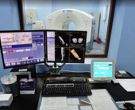
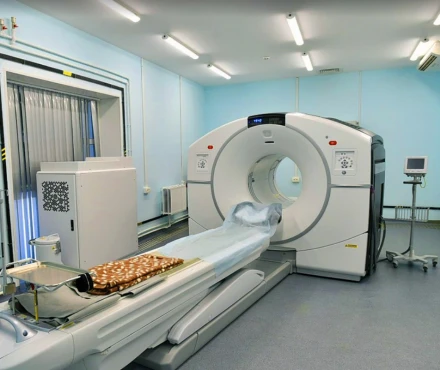
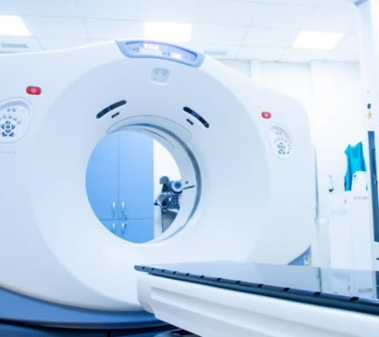
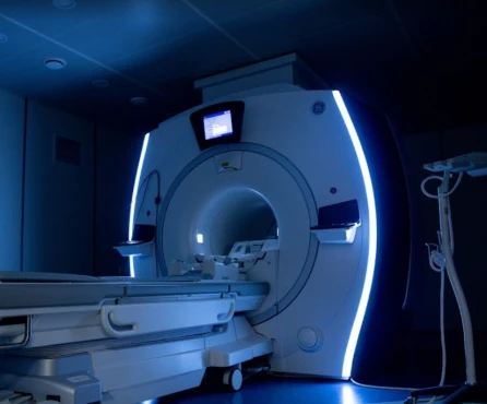
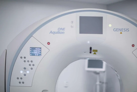
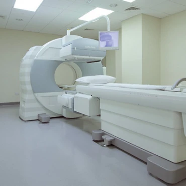
The early detection of kidney cancer is crucial for improving treatment outcomes. The diagnostic process typically involves several steps. First, a physician will take a thorough medical history and physical examination to assess any symptoms or risk factors. Next, they will order various imaging tests, such as an ultrasound, often the initial scan used to identify kidney tumors. If needed, they may also request a CT scan, which provides detailed cross-sectional images of the kidneys and is considered the gold standard for kidney cancer diagnosis. Additionally, an MRI may be used if CT scans are not recommended or to obtain more detailed images of the blood vessels and tissues.
To further investigate the condition, the healthcare team will order urine tests to check for the presence of blood or cancer cells. They will also conduct blood tests to evaluate kidney function and look for anemia or abnormal liver function signs. Sometimes, a biopsy may be performed to obtain a tissue sample for microscopic examination, providing more information about the cancer cells, such as the tumor's subtype and grade.
Kidney cancer staging
How is kidney cancer staged?
Cancer staging is used to reflect prognosis and guide treatment decisions. It describes the size and location of the tumor and whether cancer has spread to lymph nodes, organs, or other parts of the body.
The American Joint Committee on Cancer created a staging system to determine how much cancer is in the body and where it is located. This is called staging.
Based on testing, the cancer will be assigned a stage. Staging helps predict prognosis and is needed to make treatment decisions. Prognosis refers to the likely course the cancer will take. AJCC is just one type of staging system.
Information gathered during staging includes:
- The extent of the tumor: How large is the cancer? Has it grown into nearby areas?
- The spread to nearby lymph nodes: Has the cancer spread to nearby lymph nodes? If so, how many? Where?
- The spread to distant sites: Has the cancer spread to distant organs such as the lungs or liver?
- Grade of the cancer: How abnormal do the cancer cells look under a microscope?
Staging is based on a combination of information to reach a final numbered stage. Often, not all information is available at the initial evaluation. More information can be gathered as treatment begins.
Staging includes:
- Anatomic – based on the extent of cancer as defined by tumor size, lymph node status, and distant metastasis, according to the TNM scores.
- Prognostic – includes anatomic TNM plus tumor grade and other factors such as performance status. The prognostic stage also assumes that the patient is treated with the standard-of-care approaches.
Prognostic stages are divided into clinical and pathologic. Cancer staging is often done twice, before and after surgery. Staging after surgery provides more specific and accurate details about the size of the cancer, what tissue or organs may be involved, and the presence of cancer in lymph nodes.
Clinical stage is the rating given before any treatment. It is based on a physical exam, imaging tests, and possible biopsy. In kidney cancer, the clinical stage is based mainly on imaging results. These tests are done before any treatment as part of an initial diagnosis. Surgery is needed to know exactly how much cancer is in the body.
The pathologic or surgical stage is determined by examining the tissue removed during surgery. For example, pN2 indicates the cancer has spread to nearby lymph nodes. If drug therapy is given before surgery, the stage may be ypT3. The pathologic stage is based on information gained after surgery to remove all or part of the kidney and nearby lymph nodes. This provides a more accurate picture of how far the cancer has spread and helps determine treatment options after surgery. Removing the tumor tissue is an essential part of pathologic staging.
Grade
Cancer grade describes how abnormal the tumor cells appear under a microscope. Higher-grade cancers tend to grow and spread faster than lower-grade cancers. GX means the grade cannot be determined, while G1, G2, G3, and G4 indicate increasing levels of abnormality. G4 is the highest grade for renal cell carcinoma. Tumors with sarcomatoid features are also considered G4.
Numbered stages
Cancers are often described using numbered stages. These stages are based on the TNM scoring system. The stages range from 1 to 4, with four being the most advanced.
Other terminology may also be used to describe kidney cancer:
- Resectable - The tumor can be removed entirely with surgery.
- Unresectable - The tumor cannot be fully removed with surgery, as it may involve nearby blood vessels or other structures, making it unsafe to remove.
- Locoregional or locally advanced - The tumor has spread beyond the kidney to the surrounding blood vessels, tissues, organs, or lymph nodes. This would be considered a stage 3 or 4 tumor, depending on the extent of spread.
- Metastatic - The cancer has spread to other parts of the body, such as the lungs, lymph nodes, bones, liver, or brain. This is also referred to as advanced disease.
Treatment routes
What is an appropriate treatment for different kidney cancer stages?
In recent years, the treatment of kidney cancer has progressed significantly, with advancements in surgical methods, targeted therapies, and immunotherapies that aim to enhance patient outcomes.
Active surveillance
For small, slow-growing kidney tumors, especially in older patients or those with substantial health issues, active surveillance may be a suitable approach. This strategy involves regular medical imaging and close monitoring without immediate treatment. The aim is to prevent unnecessary over-treatment while keeping the option open for future intervention if the tumor starts growing [American Cancer Society, 2024].
Surgical Treatment
1. Nephrectomy is the surgical removal of the kidney, which remains the primary treatment for localized kidney cancer. There are two main types of this procedure:
- Partial nephrectomy involves removing just the tumor and a small amount of surrounding healthy tissue, preserving most of the kidney. It is commonly recommended for tumors smaller than 4 cm or in patients with a single kidney or bilateral tumors. Partial nephrectomy has a high success rate, with 5-year survival rates ranging from 75% to 95%.
- Radical nephrectomy means removing the entire kidney, along with the adrenal gland, surrounding tissue, and sometimes nearby lymph nodes. Radical nephrectomy is often used for larger or more invasive tumors. The 5-year survival rate for patients undergoing radical nephrectomy is approximately 70% to 80%.
2. Minimally invasive surgical techniques, such as laparoscopic and robotic-assisted nephrectomy, have become increasingly common in the treatment of kidney cancer. These approaches offer several benefits compared to traditional open surgery, including reduced recovery times, smaller incisions, lower blood loss, and lower rates of postoperative complications. Patients undergoing minimally invasive nephrectomy often experience a faster return to normal activities and a reduced length of hospital stay. Incorporating these advanced surgical methods has enhanced the overall outcomes and quality of life for many kidney cancer patients.
Ablation approaches
Radiofrequency Ablation (RFA) uses high-energy radio waves to heat and destroy kidney tumors. It is typically employed for patients who cannot undergo surgery due to other medical conditions. The procedure achieves local control rates of around 85%.
Cryoablation freezes and kills cancer cells. It is another option for patients unable to have surgery. The efficacy of cryoablation is similar to RFA, with local control rates of approximately 80% to 90%.
Radiation Therapy
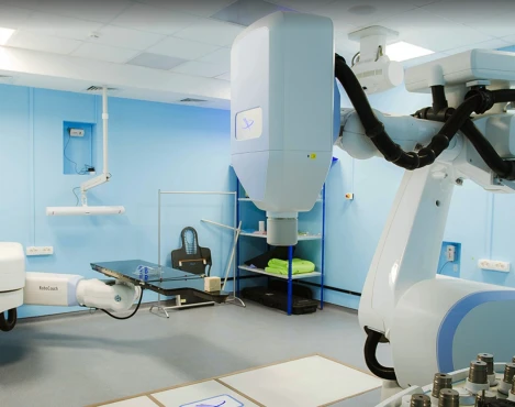
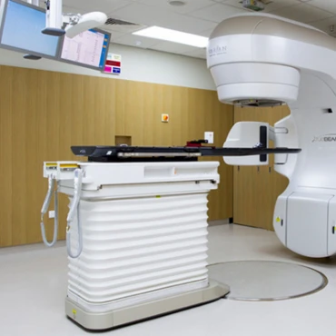
External Beam Radiation Therapy (EBRT) is primarily used palliatively in kidney cancer patients with metastases that are causing pain or other symptoms. It is not commonly used as a primary treatment due to the kidney's resistance to radiation, but it can effectively manage symptoms in advanced cases.
Systemic Therapy
1. Targeted therapy medications have transformed the treatment of advanced kidney cancer. These drugs specifically target the pathways that drive cancer cell growth and spread. Sunitinib, a tyrosine kinase inhibitor, targets multiple receptors involved in tumor growth and angiogenesis, demonstrating response rates of 30-40% and significantly improving progression-free survival. Pazopanib, another targeted therapy, inhibits blood vessel formation and offers similar efficacy to sunitinib but with a different side effect profile, providing 35-40% response rates. Axitinib is used as a second-line therapy after initial treatments fail, achieving a response rate of around 20%. Cabozantinib has also proven effective in patients with advanced kidney cancer who have exhausted prior treatment options, with a response rate of approximately 17%.
2. Immunotherapy stimulates the body's immune system to fight cancer cells. Nivolumab, a PD-1 inhibitor, has demonstrated improved overall survival in patients with advanced kidney cancer, with a response rate of about 25%. Pembrolizumab, another immunotherapy drug, is combined with axitinib, providing a 55% response rate and more prolonged progression-free survival. Furthermore, when used with nivolumab, the immunotherapy Ipilimumab has improved survival rates in advanced kidney cancer.
Chemotherapy is usually not effective against kidney cancer and is rarely used. It may be an option when other treatments fail or are not suitable.
Prognosis
How does cutting-edge science improve the lifespan and quality of life for those with the disease?
The prognosis for kidney cancer depends heavily on the stage of the disease at the time of diagnosis and the patient's response to treatment. The 5-year survival rates vary significantly based on these factors. For localized kidney cancer, the 5-year survival rate is approximately 93%. For kidney cancer that has spread to nearby regional lymph nodes or tissues, the 5-year survival rate drops to around 70%. In cases where the cancer has metastasized to distant organs, the 5-year survival rate is much lower, at only 13% [American Cancer Society, 2024].
Regular follow-up is essential for closely monitoring for potential cancer recurrence and effectively managing any long-term side effects from treatment. Recommended follow-up protocols generally involve:
- Visits every 3 to 6 months during the initial two years after completing treatment.
- Visits every 6 to 12 months for the subsequent 3 to 5 years.
- Annual visits after the first five years post-treatment.
During each follow-up appointment, the healthcare team will perform comprehensive physical exams, order necessary blood and urine tests, and use imaging methods like ultrasound, CT scans, or MRI to quickly identify any signs that cancer has returned or spread to other areas.


