Disease Types & Epidemiology
How common is liver cancer?
Primary liver cancer is a tumor that initially forms in the tissue of the liver. Different types of liver cancer exist according to the kind of cancerous cells [cancer.net].
- Hepatocellular carcinoma is the most frequent type of liver cancer. It accounts for 73% of all liver cancers. Hepatocellular carcinoma begins in hepatocytes, the principal cells of the liver, and grows invasively.
- Cholangiocarcinoma – a bile duct cancer – takes about 18% of the primary liver cancers. It develops from a thin tube – a bile duct – that extends from the liver to the small intestine.
- Angiosarcoma – rare, around 1% of primary liver cancer, arises from the blood vessels of the liver and has rapid growth.
Liver cancer represents the sixth most common cancer worldwide. In Europe, about 10 in every 1,000 men and 2 in every 1,000 women will develop liver cancer at some point in their life. Worldwide, it is much more common in South-East Asia and Western Africa. This is mainly because infection with the hepatitis B virus increases the risk of developing liver cancer and is more frequent in these areas. In the USA and Southern Europe, the hepatitis C virus is seen more frequently as a cause of liver cancer. The median age at diagnosis is between 50 and 60 years, but in Asia and Africa, it is usually between 40 and 50 years [ESMO, 2018].
Each year, about 41,210 adults in the United States and an estimated 905,677 worldwide are diagnosed with primary liver cancer.
On the other hand, tumor lesions may arise in the liver due to the metastatic process of cancers of different origins, so-called metastatic liver tumors. In this material, only primary liver cancer (hepatocellular carcinoma) is discussed. However, it is essential to mention that metastatic hepatic tumors are more prominent than primary hepatocellular or biliary tumors because the liver filtrates a blood flow coming from a large number of organs and tissues in the chest, abdomen, pelvis, and lower limbs. Metastatic stage of adenocarcinomas (tumors arising from glands in different organs) of the colorectal area, ovary, stomach, and breast in nearly 80% remain confined to the liver. Moreover, around 25% of patients diagnosed with colorectal cancer – the third most common primary malignancy worldwide – will develop liver metastases [Griscom & Wolf, 2023; Oncotarget, 2016].
Causes & Risk Factors
What is the primary issue of liver cancer?
In most patients, cancers of the liver are preceded by cirrhosis of the liver. Liver cirrhosis is a consequence of chronic liver disease, although only a limited percentage of patients with chronic liver disease will eventually develop cirrhosis. In cirrhosis, liver tissue is slowly modified at the expense of normal liver cells and consists more and more of fibrous tissue and scar tissue. In this case, the liver cells do not grow or function normally.
The exact mechanisms and reasons why liver cancer occurs are not fully understood. However, cirrhosis and its causes are the main risk factors for hepatocellular carcinoma, the primary type of liver cancer.
A risk factor increases the risk of cancer occurring but is neither necessary nor sufficient to cause cancer. It is not a cause in itself. Some people with these risk factors will never develop liver cancer, and some people without any of these risk factors will nonetheless develop liver cancer.
The main risk factors of hepatocellular carcinoma are the ones causing cirrhosis, but others not related to cirrhosis exist as well:
- Causes of liver cirrhosis:
- Chronic infection with hepatitis-B virus (HBV) or hepatitis-C virus (HCV) – if the viral diseases present for more than six months and cause a decline in the functioning of the liver. Having chronic hepatitis B infection increases the risk of developing liver cancer 100-fold, and having chronic hepatitis C infection increases the risk 17-fold. Up to 85% of individuals with hepatitis C infection develop a chronic illness; of these, approximately 30% progress to cirrhosis, and in these, 1 to 2% per year develop liver cancer.
- Long-term alcohol abuse can lead to liver cirrhosis and liver cancer.
- Inherited liver conditions include hemochromatosis (accumulation of too much absorbed iron from food) and alpha-1-antitrypsin deficiency (accumulation of abnormal protein alpha-1-antitrypsin in the liver cells).
- Non-alcoholic fatty liver disease and non-alcoholic steatohepatitis are linked to severe obesity and diabetes mellitus, and other inflammatory conditions, such as autoimmune hepatitis and intrahepatic primary inflammation.
- Gender: liver cancer is four to eight times more common in men than in women, although this is probably due to differences in behaviour that affect the risk factors described before.
- Exposure to toxic agents: anabolic steroids, aflatoxin-contaminated food (peanuts, wheat, soybeans, corn, and rice).
Clinical Manifestation & Symptoms
What signs should one anticipate while suspecting X cancer?
The main symptoms that could be related to liver cancer are the following:
- Unexplainable weight loss
- Fatigue
- Loss of appetite or feeling very full after a small meal
- Nausea or vomiting
- Fever
- An enlarged liver felt as a mass under the ribs on the right side
- An enlarged spleen felt like a mass under the ribs on the left side
- Pain in the abdomen or near the right shoulder blade
- Swelling or fluid build-up in the abdomen (ascites)
- Itching
- Yellowing of the skin and eyes (jaundice)
- Enlarged veins on the abdomen that become visible through the skin
Algorithm of diagnosis
How does the staging of liver cancer influence the best treatment strategy choice for the patient?
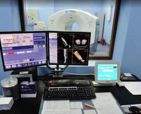
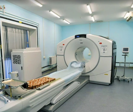
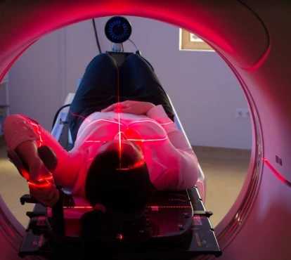
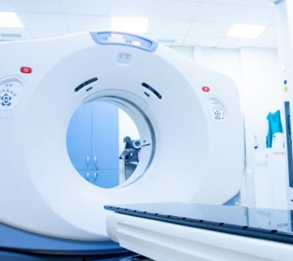
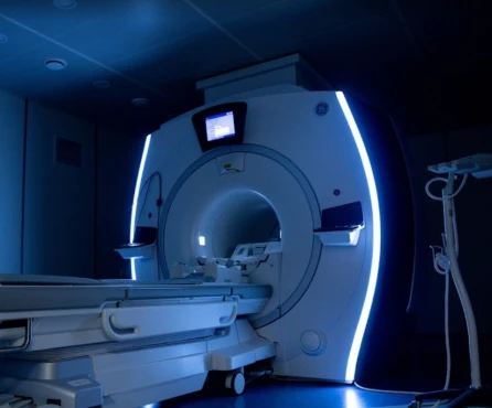
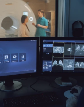
The staging of liver cancer shows the extent of the presence of a tumor in a body and the prognosis of the patient. The stage is fundamental to making the right decision about the treatment. The more advanced the stage, the worse the prognosis is. Different investigations are aimed at finding out how far the cancer has grown in and outside the liver and whether or not it has already spread to other parts of the body. Physicians perform a CT scan or an MRI scan of the abdomen to evaluate the local extent of the tumor and whether it has spread to other organs. If there is any suspicion that the cancer might have spread further away, imaging of different body parts could be performed as well, in particular, a chest CT scan and a bone scan.
Staging is usually performed twice: after clinical and radiological examination and after surgery. The removed tumor can be examined in the laboratory if surgery is performed. The results of this examination may also help stage the disease.
Since most liver cancer occurs on underlying cirrhosis, the tumor, as well as the underlying liver disease (if present), should be staged. Both determine the treatment options and the expected outcome. The first one, namely, the Child-Pugh staging system, is used to evaluate the liver cancer progression, prognosis, as well as need for transplantation in chronic liver disease.
According to the Child-Pugh system, the scoring results are divided into A, B, or C categories. ‘A’ indicates less advanced cirrhosis, and ‘C’ indicates more advanced cirrhosis. It considers the accumulation of liquid in the abdomen called ascites, the level of 2 liquid-saving proteins (called albumin and bilirubin) in the blood, how well blood clotting is still working, and the presence of encephalopathy (cognition function).
The second scale, the Barcelona Clinic Liver Cancer (BCLC) staging system, is also in active use. Its main advantage is that it identifies those patients with early cancer who may benefit from curative therapies (stage 0 and stage A), those at intermediate (stage B) or advanced stage (stage C) who may benefit from life-prolonging treatments, and those with very limited life expectancy (stage D). The BCLC system is based on the size and the number of (the) tumor (s) in the liver, invasion of blood vessels by the tumor, cancer spread outside the liver, blood pressure in the vein going to the liver, level of bilirubin in the blood, Child-Pugh score and performance status.
The blood pressure in the vein going to the liver (called portal vein) can be raised when the liver does not let the blood pass through quickly because of a changed consistency. Bilirubin is a protein usually excreted by the liver into the bile. However, when the liver function is impaired, it can also be seen in the blood. The Child-Pugh score has been described before and considers the accumulation of liquid in the abdomen (ascites), the level of albumin and bilirubin in the blood, how well blood clotting works, and the presence of encephalopathy. The performance status has been described in the previous section. It evaluates the patient’s physical abilities by giving a score from 0 for a fully active patient to 4 for a patient who is completely disabled due to their disease.
Because the BCLC includes so many factors, it is found to give the best prediction of the prognosis for the patient who has cirrhosis and liver cancer and to be very useful in planning the treatment.
Treatment routes
What is an appropriate treatment for different stages of liver cancer?
The treatment plan for hepatocellular carcinoma (HCC) mainly depends on the stage according to the BCLC staging system.
Treatment protocol for patients with stage 0 and stage A of HCC according to BCLC
- Tumor’s surgical resection for:
- Patients without cirrhosis and for whom a sufficient part of the liver can be preserved;
- Patients with BCLC stage 0 or A whose performance status allows them to undergo surgery.
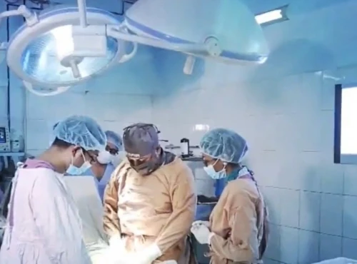
The resection of the tumor consists in removing the part of the liver which contains the lesion. This type of surgery is called a partial hepatectomy, and it can only be performed in patients without cirrhosis or with limited cirrhosis since the liver still functions correctly in these patients. The remaining part of the liver will take over the liver function. After surgery, the resected part will be examined by a pathologist in the laboratory. The pathologist will check whether the whole tumor has been removed by analyzing if the lump is totally surrounded by normal tissue. This is reported either as negative margins of resection, meaning that it is very likely that the whole tumor has been removed, or as positive margins of resection, meaning that it is very likely that the entire tumor has not been removed. If margins are negative, it is a sign of a better prognosis.
- Liver transplantation. When resection of the tumor is not possible, liver transplantation should be considered either when there is a single tumor of less than 5 cm in diameter or when there are 2 to 3 tumors, each measuring less than 3 cm in diameter. However, Patients who suffer from liver cirrhosis caused by alcohol abuse and are still using alcohol, or patients who have a poor prognosis due to the characteristics of their cancer or due to other concurrent diseases, will not be considered for transplantation.
Liver transplantation is an operation under a general anesthetic that usually takes 6 to 10 hours. During this time, the surgeons will first make an incision shaped like a boomerang on the upper part of the abdomen and remove the patient’s old liver, leaving portions of the major recipient blood vessels in place. The new liver will then be inserted and attached to these blood vessels and the patient’s bile ducts.
Owing to organ shortage, liver transplant candidates are confronted with long waiting times, which must not delay the discussion about an alternative effective treatment. In the case of a long-anticipated waiting time (> six months), patients may be offered resection, local ablation, or transarterial chemoembolization to minimize the risk of tumor progression and to provide a ‘bridge’ to transplant.
- Local ablation methods. Local ablation aims to destroy cancer cells by targeting them with chemical or physical means. The two main local ablation methods are radiofrequency ablation and percutaneous ethanol injection, which will be described further. While these techniques effectively destroy small tumors, they, unfortunately, do not prevent the occurrence of new lesions in the surrounding cirrhotic liver tissue.
Two methods show similar results for BCLC stage 0 tumors:
- Radiofrequency ablation uses high-energy radio waves to destroy cancer cells. It gives better results in control of tumor growth in tumors larger than 2 cm in diameter.
- Percutaneous ethanol injection uses ethanol (concentrated alcohol) to scorch the tumor. The ethanol is injected directly into the tumor through the skin. Sometimes, an ultrasound or CT scan guides the needle right into the tumor.
Treatment protocol for patients with stage B of HCC according to BCLC
- Transarterial chemoembolization (TACE) is an injection of small degradable particles (lipoidol) and anticancer drugs (doxorubicin, cisplatin, mitomycin) directly in the artery that supplies the liver with blood under X-ray control. Tumor cells preferentially will absorb lipoidol and the medication at the same time.
- TACE with radiopharmaceutical drugs, which are part of radiation therapy with radioactive substances Iodine 131 or Yttrium 90. They are released in microscopic balls via a small tube into the main artery going to the liver, blocking the blood supply to the tumor and emitting radiation that destroys the tumor cells surrounding them. An advantage is that it can be used regardless of how numerous or significant the nodules in the liver are, and it can also be used to treat tumors possibly undetected.
- Targeted therapy with sorafenib, which blocks the tumor growth, is used in case of HCC progression despite TACE.
Treatment protocol for patients with stage C of HCC according to BCLC
- Systemic therapy with:
- Sorafenib, which extends the survival rate for an average of 2.8 months for patients with Child-Pugh grade A;
- Chemotherapy – less selective and has more side effects such as anemia – with capecitabine, oxaliplatin, or gemcitabine in combinations. The chemotherapy shows an oncostatic impact, meaning it blocks the tumor progression but does not eliminate the existing lesions.
- Radiotherapy such as:
- Radioembolization with yttrium-90 microspheres for patients who suffer from blood clots blocking a branch of one of the main veins of the liver, called portal vein thrombosis;
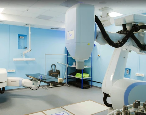
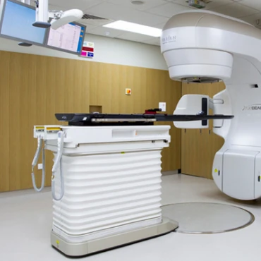
- External radiotherapy by three-dimensional conformal radiotherapy (3D-CRT). Radiations are produced by a device outside the body and then directed toward the tumor. It is called 3D-conformational because, unlike classic external radiotherapy techniques, a computer calculates the exact direction and shape of the radiation beams. This helps direct them precisely to the tumor and leave as many normal liver cells as possible unharmed.
Treatment protocol for patients with stage D of HCC according to BCLC - only symptomatic treatment to control pain and nausea, combined with biliary duct stenting to ensure a free passage of the bile into the intestines to prevent rapid inner intoxication.
Prognosis
How does cutting-edge science improve the lifespan and quality of life for those with liver cancer?
According to the American Cancer Society, the five-year relative survival rate for early liver cancer (HCC) is 37%, with intermediate-advanced cancer – 14% and with terminal HCC - 4%. Regardless of the stage, the survival rates are higher for surgically resectable cases. For people with early-stage liver cancers who have a liver transplant, the 5-year survival rate is in the range of 60% to 70% [cancer.org].


