Disease Types & Epidemiology
How common are the mediastinal tumors?
Mediastinal growths are abnormal lesions found in the mediastinum, the central region of the chest cavity located between the lungs. This area contains vital structures such as the heart, thymus, esophagus, trachea, and major blood vessels. These growths can be either primary – originating in the mediastinum or secondary – spreading from tumors of other body parts. They are relatively uncommon and comprise approximately 3% of all thoracic tumors. The occurrence varies according to age group and tumor type; primary mediastinal growths are more prevalent among younger patients, whereas metastatic tumors are seen more frequently in older adults. Altogether, primary and metastatic tumors of mediastinum amount to around 7,000 cases per year [ESMO, 2019].
The mediastinum is generally divided into three main parts: anterior (front), posterior (back), and middle. It is bordered by the thoracic inlet at the top, the diaphragm at the bottom, the spine in the back, the sternum in front, and pleural spaces on each side. The aggressiveness of cancer and how severe its signs and symptoms depend on how it behaves within this visceral network. Various types of cancers can occur in the mediastinum. The prognosis for each type depends on its behavior, tendency to spread or invade other areas, and resistance to treatment. Therefore, all tumors found in this area can be categorized based on their location as either front-, mid-, or back-located.
1. Anterior mediastinum tumors are enclosed by the pericardium at the back, pleural sacs on the sides, and the sternum in front.
- Thymomas and thymic carcinomas. Thymomas are the most frequent primary tumors found in the front part of the mediastinum in adults are thymomas. These lesions are derived from thymic epithelial cells and tend to develop slowly, often linked to myasthenia gravis, an autoimmune neuromuscular condition. However, 10% of thymic tumors are malignant and called thymic carcinomas, which may be highly aggressive, early-metastasizing cancers. These pathologies account for approximately 20-30% of all tumors in the mediastinal region [University of Illinois. 2024].
- Lymphomas. Both Hodgkin's (12% of cases) and non-Hodgkin's lymphomas (13%) may develop in the front part of the mediastinum, especially in younger individuals. They make up around 25% of tumors found in this area.
Primary lymphomas that manifest as a disease in the mediastinum include primary mediastinal large B-cell lymphoma, Nodular Sclerosing Hodgkin's disease, marginal zone lymphoma of mucosa-associated lymphoid tissue, and also rarer T-lymphoblastic leukemia or lymphoma, anaplastic large cell lymphoma, and other T-, B-, and NK-cell types. In contrast, secondary lymphomas originating elsewhere in the body and spreading to the mediastinum are more prevalent than primary mediastinal lymphomas [European Respiratory Review, 2021].
- Germ Cell Tumors. Reproductive cell-derived tumors such as teratomas, seminomas, and non-seminomatous germ cells are the source. They comprise roughly 20% of anterior mediastinal masses, with benign teratomas accounting for 10% and malignant germ-cell tumors accounting for another 10%.
- Neuroendocrine tumors. Primary neuroendocrine tumors (NETs) in the mediastinum are rare, making up approximately 5% of all thymic and mediastinal tumors worldwide. The majority of thoracic NETs develop in the front part of the chest cavity within the thymus, comprising about 15% of anterior mediastinal tumors.
2. Middle mediastinum tumors are bounded by the pericardial and bilateral pleural sacks both at the front and from the back.
- Parathyroid adenoma. Several tumors have the potential to metastasize to the middle mediastinum, including parathyroid adenoma. Primary hyperparathyroidism impacts up to 0.5% of the population, and its incidence increases after 50 years of age. While a quarter of cases involve ectopic foci, only about 2% are located in the mediastinum, with even fewer found in the middle portion [Razzak et al., 2016].
- Bronchogenic Cysts. Lesions that originate from the bronchial tree at birth are usually non-cancerous. Surgical removal is necessary due to the potential for complications such as infection, rupture, or formation of passages into nearby structures, as well as the risk of the lesion returning if not completely removed.
- Known as pericardial cysts, benign fluid-filled sacs are typically found near the heart in the mediastinum. While they can appear anywhere in this area, they are most commonly located in the cardiophrenic angle, predominantly on the right side.
- Lymphadenopathy is an enlargement of the lymph nodes due to infections, cancers, or other inflammatory issues. This term describes the abnormal increase in size and irregular shape of mediastinal nodes, along with changes in their density. According to commonly accepted standards, mediastinal lymphadenopathy is characterized by a node with a short axis measuring over 10 mm.
3. Posterior Mediastinum tumors are located between pericardium at the front and spine from the back:
- Neurogenic tumors account for more than 60% of the masses discovered in the back part of the chest, typically affecting children and often growing to a significant size before causing symptoms. Around 30% of these tumors are cancerous. They mainly fall into nerve sheath neoplasms (such as schwannomas and neurofibromas) and ganglion cell neoplasms (including neuroblastomas and ganglioneuroblastomas).
- Esophageal Tumors. The lymphatic network surrounding the esophagus is extensive. Tumors in the upper or middle part of the esophagus often spread to the thoracic nodes, with paraesophageal and paratracheal nodes being frequently affected. Nodal metastasis is identified by nodal enlargement exceeding 1 cm in diameter [University of Illinois. 2024].
Causes & Risk Factors
What is the primary issue of mediastinal tumors?
The exact cause of mediastinal tumors often remains unclear. However, certain risk factors have been identified:
- Genetic Predisposition. Some patients have a genetic predisposition to developing thymomas (for example, GTF2I gene mutation in 74%-82% of cases) and germ cell tumors (TP53 / KRAS oncomarkers in 18%-46%) [University of Antwerp, 2022; University of British Columbia, 2022].
- Autoimmune Conditions. According to Myasthenia Gravis Foundation, 20% of thymomas are associated with autoimmune diseases [Myasthenia Gravis Foundation, 2024].
- Previous Radiation Exposure. Prior chest radiation therapy can elevate the likelihood of developing tumors in the mediastinal area, especially lymphomas and sarcomas.
- Congenital Factors. Conditions like neurofibromatosis are associated with the formation of neurogenic tumors in the back part of the mediastinum.
Clinical Manifestation & Symptoms
What signs should one anticipate while suspecting a mediastinal tumor?
The signs of mediastinal tumors can vary widely, depending on the tumor's size, location, and type. Common symptoms include chest discomfort that may worsen with breathing or coughing, persistent and unproductive coughing, difficulty swallowing due to compression of the esophagus, hoarseness caused by nerve compression in the throat area, shortness of breath from airway or lung tissue compression, non-specific symptoms like weight loss and fatigue which could be linked to malignancy. There might also be swelling of the face, neck, and upper limbs due to obstruction of a major vein called superior vena cava.
Diagnostic Route & Screening
When, where, and how should mediastinal tumors be detected?
Mediastinal cancer is usually conclusively diagnosed through a procedure called mediastinoscopy with biopsy, which involves collecting cells from the mediastinum to identify the type of mass present. This test is commonly performed under general anesthesia.
However, the diagnosis of mediastinal tumors typically involves a combination of imaging studies and tissue sampling, such as:
Imaging methods:
- Chest X-ray is frequently used as the initial imaging technique to identify a mediastinal mass.
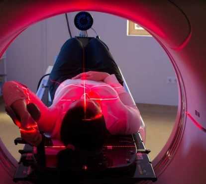
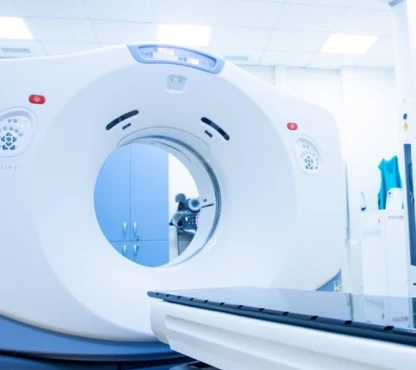
- Computed Tomography (CT) scan offers comprehensive cross-sectional images of the chest, aiding in determining the size, location, and potential impact on neighboring tumor structures.
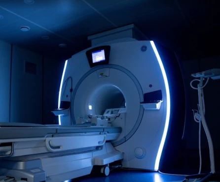
- Magnetic Resonance Imaging (MRI) is valuable for assessing soft tissue involvement and vascular structures.
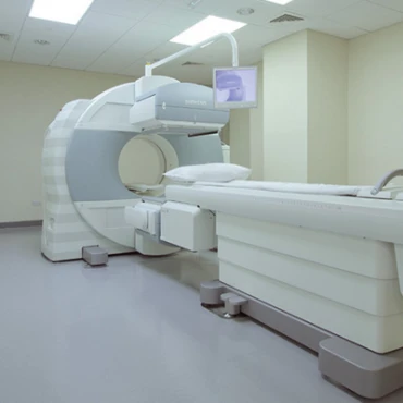
- Positron Emission Tomography (PET) scan assists in evaluating metabolic activity and distinguishing between benign and malignant masses.
The biopsy technique that is used for mediastinal tumor diagnosis is performed through mediastinoscopy – a surgical procedure employed to obtain tissue samples from the mediastinal lesion for histopathological examination. The piece of tumor tissue can be extracted from the patient via two approaches:
-
Fine Needle Aspiration (FNA) is a minimally invasive approach for extracting cells for analysis.
-
Core Needle Biopsy yields a larger tissue sample compared to FNA.
Laboratory examinations include blood tests involving tumor markers like alpha-fetoprotein, beta-human chorionic gonadotropin (β-hCG) for germ cell tumors, and lactate dehydrogenase (LDH) for lymphomas.
Introduction & Principles Mediastinal Tumors Staging
Mediastinal tumors are a diverse group of neoplasms located in the mediastinum, the area between the lungs that houses vital structures such as the heart, trachea, and esophagus. Treatment protocols involve tumor staging.
Mediastinal tumors are a diverse group of neoplasms located in the mediastinum, the area between the lungs that houses vital structures such as the heart, trachea, and esophagus. Treatment protocols involve tumor staging.
Mediastinal tumors are categorized based on the TNM staging system, which evaluates the spread of the tumor by assessing the primary tumor (T), regional lymph nodes (N), and distant metastasis (M). This classification system is crucial in guiding treatment approaches and forecasting prognosis. Thus, early-stage (I-II) and advanced-stage (III-IV) mediastinal tumors have key distinguishing according to the TNM classification.
Early-stage mediastinal tumors (stages I-II)
Stage I
- T1: The tumor is confined to the mediastinum and does not invade surrounding structures.
- N0: No regional lymph node involvement.
- M0: No distant metastasis.
Stage II
- T2: The tumor extends into adjacent structures within the mediastinum, such as the pericardium, but not beyond.
- N0: No regional lymph node involvement.
- M0: No distant metastasis.
Mediastinal tumors in the early stages are usually confined and have not metastasized to lymph nodes or other organs. Because of this limited spread, surgical removal is often used as the primary form of treatment. At these initial stages, the tumors tend to be smaller and have not infiltrated vital organs.
Advanced-stage mediastinal tumors (stages III-IV)
Stage III
- T3: Tumor invades nearby structures, such as the chest wall, diaphragm, or pleura.
- N1/N2: Regional lymph node involvement, indicating the spread to nearby lymph nodes on the same side of the diaphragm (N1) or both sides (N2).
- M0: No distant metastasis.
Stage IV
- T4: Tumor invades more distant structures, such as the heart, great vessels, trachea, esophagus, or vertebral body.
- N3: More extensive lymph node involvement, including supraclavicular or scalene nodes.
- M1: Distant metastasis is present, indicating that it has spread to distant organs.
Advanced-stage mediastinal tumors are known for extensive local invasion, significant lymph node involvement, and potential distant metastasis. Treatment usually involves a combination of surgery, chemotherapy, and radiation therapy.
Treatment routes
What is an appropriate treatment for mediastinal tumors at different stages?
- Anterior Mediastinum Tumors
1.1 Thymomas [Zhang et al., 2023]
Early-stage (stage I-II)
- First-line therapy: Surgery is the main treatment option. A successful resection has been linked to a solid 5-year survival rate of around 80-90%.
- Second-line therapy: Post-surgery radiation therapy is advised if the surgical margins are positive or if there is an invasion of the capsule. Using radiation therapy alongside surgery can significantly decrease the rates of local recurrence.
Advanced-stage (stage III-IV)
- First-line therapy: Combination therapy using cisplatin, doxorubicin, and cyclophosphamide (the CAP regimen) is the established treatment for advanced thymomas. The response rate to this combination is approximately 50-70%.
- Second-line therapy: For cases that are difficult to treat, alternative options involve pemetrexed and carboplatin, demonstrating a moderate response rate of approximately 30%.
1.2. Thymic carcinomas [Tartarone et al., 2023]
Early-stage (stage I-II)
- First-line therapy: Surgery followed by radiation therapy. The five-year survival rate after treatment is around 50%-60%.
- Second-line therapy: Chemotherapy using treatments such as paclitaxel and carboplatin is utilized when surgery fails to cure completely.
Advanced-stage (stage III-IV)
- First-line therapy: Chemotherapy using schedules like CAP or carboplatin and paclitaxel demonstrates response rates similar to those observed in thymomas.
- Second-line therapy: Targeted treatments such as sunitinib have been studied for refractory thymic carcinomas, showing response rates of approximately 20%.
1.3. Lymphomas [University of Illinois, 2024]
Early-stage (stage I-II)
- First-line therapy: Chemotherapy treatments such as CHOP (cyclophosphamide, doxorubicin, vincristine, prednisone) combined with radiation therapy have shown a 5-year survival rate of approximately 70-80%.
- Second-line therapy: Salvage chemotherapy with ICE (ifosfamide, carboplatin, etoposide) is employed in situations of relapse.
Advanced-stage (stage III-IV)
- First-line therapy: Intensive treatment plans like EPOCH (etoposide, prednisone, vincristine, cyclophosphamide, and doxorubicin) are commonly used and typically result in response rates of about 60%.
- Second-line therapy: High-dose chemotherapy with subsequent autologous stem cell transplantation can be considered for cases that do not respond to standard treatments.
1.4. Germ cell tumors
Early-stage (stage I-II)
- First-line therapy: Surgery is the primary treatment, and early-stage germ cell tumors have a 5-year survival rate of over 90%.
- Second-line therapy: If residual masses are found, the option of post-operative treatment with BEP chemotherapy (bleomycin, etoposide, cisplatin) should be considered.
Advanced-stage (stage III-IV)
- First-line therapy: Chemotherapy with the BEP protocol can yield around 70-80% response rates.
- Second-line therapy: High-dose chemotherapy and autologous stem cell transplant for cases that do not respond to conventional treatment.
1.5. Neuroendocrine tumors
Early-stage (stage I-II)
- First-line therapy: Surgery is the most common form of treatment. Early-stage neuroendocrine tumors generally have a 5-year survival rate of around 80-90%.
- Second-line therapy: Post-surgery chemotherapy using drugs such as octreotide to manage symptoms.
Advanced-stage (stage III-IV)
- First-line therapy: Chemotherapy treatments such as streptozocin, 5-FU, and doxorubicin are associated with approximately 30-40% response rates.
- Second-line therapy: Targeted treatments such as everolimus or sunitinib may effectively treat cases that have not responded to other therapies.
2. Middle mediastinum tumors
2.1. Parathyroid adenoma: early-stage is prevalent. Thus, surgical removal is the primary treatment with a high cure rate of around 90%.
2.2. Bronchogenic cysts are typically cured via surgical resection in early- and advanced stages.
2.3. Pericardial cysts, most of the time, resolve on their own. However, if symptoms such as chest pain or difficulty breathing occur, surgery may be necessary. Pleuropericardial cysts can pose risks such as bleeding, atrial fibrillation, and cardiac tamponade. Patients without symptoms can receive conservative treatment with regular monitoring.
2.4. Lymphadenopathy’s treatment approach depends on the underlying cause, often managed therapeutically, not surgically. Thoracic lymphadenopathies can have inflammatory or neoplastic causes. Infections like tuberculosis and fungal infections such as histoplasmosis frequently lead to mediastinal lymph node enlargement. Additionally, sarcoidosis, drug reactions, heart failure, silicosis, and Castleman disease are other conditions that can cause thoracic node enlargement.
3. Posterior Mediastinum tumors
3.1. Neurogenic tumors are treated primarily by surgical operation on all stages of the disease. A roughly 90% survival rate over five years for non-cancerous growths has been observed. For malignant tumors, chemotherapy or radiation may be added.
3.2. Esophageal mediastinal tumors
Early-stage (stage I-II)
First-line therapy: Surgery to remove the tumor is associated with a 50% survival rate for early-stage esophageal tumors over five years from diagnosis.
Advanced-stage (stage III-IV)
First-Line Therapy: Chemotherapy using drugs such as cisplatin and 5-FU has shown response rates of around 30-40%.
A few approaches, such as immunotherapy and targeted therapies, are currently under investigation to treat refractory mediastinal tumors. Clinical trials have indicated the potential effectiveness of drugs like pembrolizumab and nivolumab, providing optimism for patients with few alternatives.
Follow-up
How does cutting-edge science improve the lifespan and quality of life for those with the disease?
The prognosis for mediastinal tumors differs greatly depending on the tumor type when it was diagnosed, and how it responds to treatment. For example, thymomas and germ cell tumors in the early stages often have a survival rate of over 90%, while advanced-stage thymic carcinomas and neuroendocrine tumors typically have worse results.
Regular follow-up is crucial for detecting recurrences and managing long-term side effects of treatment. This usually involves a follow-up protocol that includes:
Initial follow-Up. Every 3 to 6 months during the initial two years following treatment, which involves both physical examinations and imaging tests.
Long-term follow-up. After the initial two years, the focus will shift to addressing survivorship concerns and conducting screenings for secondary malignancies yearly.
Prognosis
How does cutting-edge science improve the lifespan and quality of life for those with the disease?
The prognosis for patients with mediastinal tumors varies widely based on the type and stage of the tumor at diagnosis:
- Thymomas generally have a favorable prognosis with a 5-year survival rate of about 70-90%, mainly if detected early and surgically resected.
- Lymphomas: prognosis depends on the specific subtype and response to chemotherapy and radiation. Hodgkin lymphoma has a high cure rate with a 5-year survival rate of about 85-90% in its early stages.
- Germ Cell Tumors often have an excellent prognosis with a high cure rate following surgery and chemotherapy. Benign teratomas have a nearly 100% 5-year survival rate, while malignant germ cell tumors have a 5-year survival rate of 60-80% when treated appropriately
- Neurogenic Tumors: prognosis is generally good for benign tumors that can be completely resected. Malignant neurogenic tumors, however, can be more challenging with a 5-year survival rate of 30-50%.


