Disease types, risk factors & epidemiology
How common is the disease?
Gastrointestinal stromal tumors (GISTs) are a rare type of tumor found primarily in the stomach (in 60% of GIST cases) and small intestine (30%). These tumors develop from the interstitial cells of Cajal, which are part of the intestine's autonomic nervous system and help regulate its muscular contractions. GISTs are distinct from other gastrointestinal tumors due to their unique genetic characteristics, often involving mutations in the KIT (85% of GIST) or PDGFRA (7%) genes.
GISTs are the most common mesenchymal tumors of the digestive tract, though they remain relatively uncommon. The annual incidence is estimated at 10-20 cases per million people in the United States. At the exact moment, it is estimated that there are about 900 new cases of GIST diagnosed per year in the UK. While most GISTs occur sporadically, a small percentage are associated with genetic syndromes like Neurofibromatosis type 1 and familial GIST syndromes. These tumors can develop at any age but are most frequently diagnosed in individuals between 50 and 70 years old [Spanish Group for Sarcoma Research, 2023].
Clinical Manifestation & Symptoms
What signs should one anticipate while suspecting gastrointestinal stromal tumors?
The symptoms of gastrointestinal stromal tumors can vary depending on the size and location of the cancer. Gastric GISTs may cause abdominal discomfort, gastrointestinal bleeding, nausea, and vomiting. Small intestinal GISTs can lead to abdominal pain, intestinal blockage, and gastrointestinal bleeding. Meanwhile, colorectal and esophageal GISTs can result in swallowing difficulties, rectal bleeding, and alterations in bowel movements.
Diagnostic Route
When, where, and how should gastrointestinal stromal tumors be detected?
The diagnosis of GISTs involves a combination of imaging tests, endoscopic procedures, and microscopic tissue analysis:
1. Imaging tests:
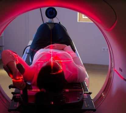
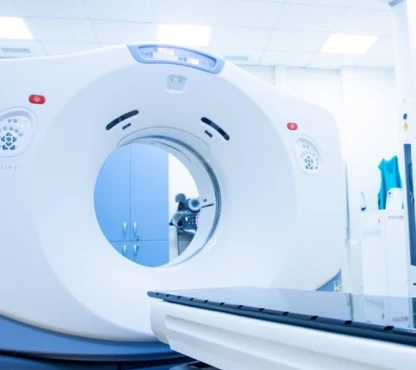
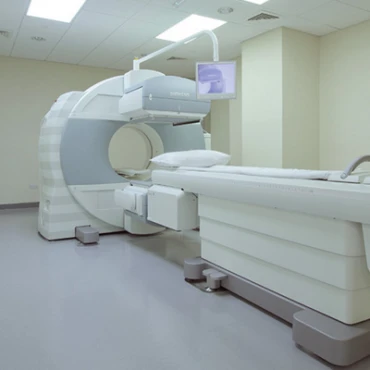
- CT Scan visualizes the tumor and assesses its size and extent.
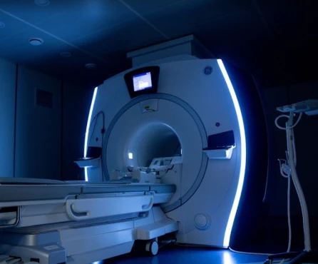
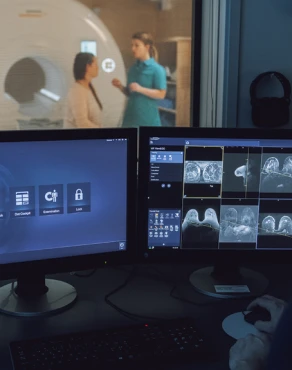
- MRI may be used for further evaluation, particularly for rectal GISTs.
- PET scan helps assess the tumor's metabolic activity and identify any metastases.
2. Endoscopic Procedures:
- Endoscopic ultrasound helps evaluate tumors in the stomach and small intestine.
- Endoscopic biopsy obtains tissue samples for microscopic examination.
3. Microscopic tissue analysis, such as immunohistochemistry with staining for KIT (CD117) and DOG1 proteins, helps confirm the diagnosis of GIST.
Treatment Approaches [ESMO, 2022]
What are the options for managing gastrointestinal stromal tumors?
Surgery
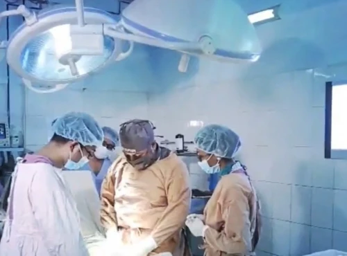
If the tumor is small, easy to get at, and has not spread, surgery is the usual treatment. This may be possible laparoscopically (by keyhole surgery), but open surgery may be needed. The response rates for surgical resection of early-stage GIST are approximately 60-70%.
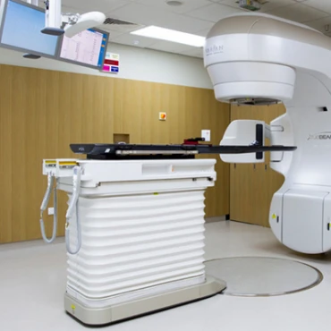
Targeted therapy (imatinib, sunitinib, regorafenib) is an adjuvant (imatinib for small GIST before and after the surgery) or independent option for advanced disease. Thus, targeted therapy's average success frequency numbers are 50-60% for imatinib, 20-30% for sunitinib, and about 15% for regorafenib.
If targeted therapy fails, chemotherapy with doxorubicin and ifosfamide gives around 10-15% to cure the GIST.
Prognosis & Follow-up
How does cutting-edge science improve the lifespan and quality of life for those with gastrointestinal stromal tumors?
The prognosis for GISTs can vary significantly depending on several key factors. Patients with localized GISTs that have been entirely removed through surgery generally have a favorable 5-year survival rate of around 70-80%. However, for those with advanced GISTs that have spread to other parts of the body, the 5-year survival rate drops to around 50%, though targeted therapies have helped improve outcomes in these more complex cases.
Ongoing medical monitoring is essential for effectively managing GISTs. Patients should attend follow-up visits every 3 to 6 months in the initial period. These appointments will involve physical exams, imaging tests, and vigilance for any signs of tumor recurrence or further progression. As time passes and the condition remains stable, the frequency of these follow-up visits may decrease to once every 6 to 12 months, usually after the first 2-3 years.


