Disease Types & Epidemiology
How common is the disease?
Head and neck cancers are a group of cancers that originate from the upper aerodigestive tract (lips, tongue, mouth, throat, and larynx or voice box), the salivary glands, the nasopharynx (the area that connects the nose and the upper part of the throat) or the sinuses and nasal cavity. Almost all cancers in these areas are squamous cell carcinomas (SCCHN), meaning they arise from the mucosal linings of these body parts.
These types of cancer make up about 4% of all cancer cases in the United States, with around 58,450 new diagnoses and 12,230 deaths expected in 2024 [American Cancer Society data, 2024].
The location most frequently affected is the oral cavity, accounting for 41% of all head and neck cancers, followed by pharynx and larynx cancers with 22% and 24%, respectively.
Other types of tumors, like epithelial tumors of nasal cavities, nasopharynx, eye and adnexa, and middle ears, occur in less than five people per million. These cancer types are categorized as rare cancers of the head and neck.
Causes & Risk Factors
What is the primary issue of squamous cell carcinoma of the head and neck?
Tobacco consumption and alcohol drinking have been linked to more than 70% of head and neck cancers. Some other risk factors have been identified as well.
Tobacco consumption. The risk is directly linked to the time and quantity of tobacco consumption. It has been found, however, that the risk decreases in time after quitting its use. Secondhand smoking (passive smoking) also increases the risk. Not only tobacco smoking but the consumption of smokeless tobacco, such as chewing tobacco and snuff, has been associated with oral cancer.
Alcohol. Alcohol consumption has been associated with the majority of cases of head and neck cancer. The risk associated with alcohol drinking increases in time and proportionally to the quantities of alcohol consumed. Heavy drinkers are at higher risk, 5−fold for oral cancer and 7−fold for pharyngeal cancers.
Human papillomaviruses (HPV). Evidence of infection with HPV, particularly HPV16, has been found in cancers of the oropharynx, much less in the oral cavity and larynx. Moreover, sexual behaviors are correlated to head and neck cancers, such as earlier age at sexual debut and multiple sexual partners.
Having a first−degree relative (parents, siblings, or children) affected and low socioeconomic status have also been correlated with head and neck cancers. However, they could only be reflecting the variability of exposure to alcohol and tobacco consumption.
Other important risk factors are a diet high in animal fats and low in fresh fruit for all types of head and neck cancer, prolonged exposure to sunlight for lip cancer; gastro−oesophageal reflux disease for larynx and pharynx cancers, radiation exposure for salivary glands cancer and yerba mate drinking for oral cavity cancers. There are also some pre−cancerous conditions, such as white and red patches (leukoplakia and erythroplakia, respectively) associated with tobacco use or other conditions that increase the risk of developing cancer in the mouth.
Clinical Manifestation & Symptoms
What signs should one anticipate while suspecting squamous cell carcinoma of the head and neck?
Head and neck cancers can be suspected with the appearance of symptoms depending on the cancer's specific location. Lump in the neck, tongue sore, bleeding area, white or red patch in the mouth, pain in the throat, painful swallowing, persistent hoarseness, blocked nose on one side, and/or bloody discharge from the nose are symptoms that when persistent for more than three weeks, should be investigated by a doctor.
For example, there might be sores that do not heal in the oral cavity and persistent mouth pain. In the pharynx, symptoms could include difficulty swallowing, throat pain, or a lump in the neck. If it is the larynx, hoarseness, or trouble breathing, a persistent cough might occur. Tumors in the sinuses and nasal cavity could result in blocked sinuses, chronic sinus infections, nose bleeds, or headaches. At the same time, those affecting salivary glands may cause swelling under the chin or around the jaw, numbness, or paralysis of facial muscles.
Diagnostic Route
When, where, and how should the head and neck squamous cell carcinoma be detected?
Diagnosis typically involves a combination of physical examinations, imaging studies, endoscopy, and biopsy:
- Physical Examination may include visual inspection of the mouth, nose, and neck, using a light and a mirror for a clearer view. Observation and palpation of the lips, cheeks, gums, and neck are performed to investigate for lumps or other abnormalities.
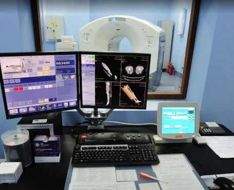
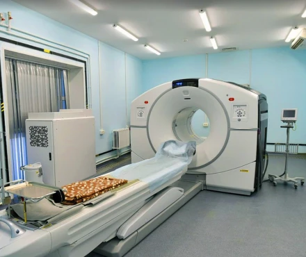
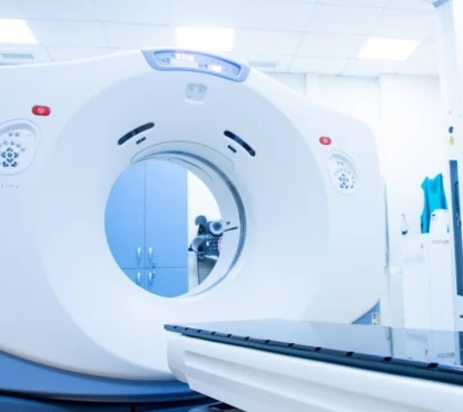
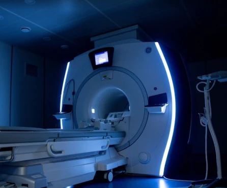
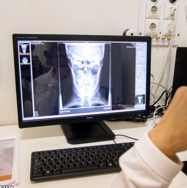
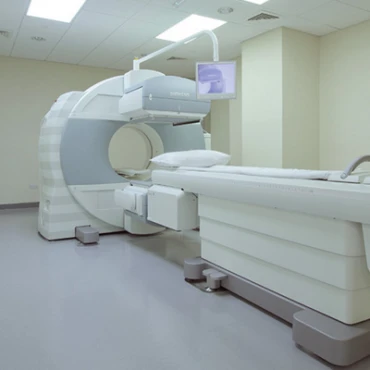
- Imaging - CT, MRI, and PET scans - are used to assess the extent of the disease.
A CT scan can show soft tissues, including lymph nodes, bony structures, and blood vessels, at the same time. However, MRI has a better resolution when compared to picture details of soft tissues. Hence, MRI is the preferable staging procedure for every tumor subsite except laryngeal and hypopharyngeal cancers.
A chest X-ray or PET scan is recommended to evaluate the eventual presence of metastases in the lung or a primary tumor in the lung. In that regard, a chest CT scan may be performed for more extensive tumors.
- Endoscopy. While the oral cavity and oral pharynx may be inspected directly, visualizing the nasopharynx, hypopharynx, and larynx requires the use of indirect mirror laryngoscopy and/or endoscopy, a procedure to examine the areas inside the body using a thin, lighted and flexible tube called endoscope.
- Biopsy. A tissue sample is taken for histopathological examination to confirm the diagnosis. This action can be done via endoscopy and, depending on tumor location, simply by opening the mouth or, in some cases, by taking a sample from an enlarged lymph node in the neck.
Doctors use staging to assess the cancer's extension and the patient's prognosis. The TNM staging system is commonly used. The combination of T (size of the tumor and invasion of nearby tissue), N (involvement of lymph nodes), and M (metastasis or spread of the cancer to other body parts) will classify the cancer into one of the stages explained later.
The stage is fundamental to making the right decision about the treatment. The lower the stage, the better the prognosis. Staging is usually performed twice: after clinical and radiological examination and after surgery. If surgery is performed, staging may also be influenced by the laboratory examination of the removed tumor. Staging is very particular for each cancer location as the structures affected differ. However, there is a general principle of SCCHN diagnosis.
Stage I
- Tumor size and infiltration: 2 cm in diameter or less with no infiltration of contiguous tissues.
- Lymph nodes involved: No.
- Distant organs involved: No.
Stage II
- Tumor size and infiltration: Tumour of more than 2 cm but no more than 4 cm or already involving neighboring sites.
- Lymph nodes involved: No.
- Distant organs involved: No.
Stage III
- Tumor size and infiltration: Tumour of more than 4 cm.
OR
- Lymph nodes involved: Yes, and 3 cm maximum.
- Distant organs involved: No.
Stage IVA
- Tumor size and infiltration: Any size and infiltration.
- Lymph nodes involved: Yes, and between 3 and 6 cm.
- Distant organs involved: No.
Stage IVB
- Tumor size and infiltration: The tumor invades the space in front of the spine in the neck, the carotid artery, or structures in the area between the lungs called mediastinum, such as the trachea and esophagus.
OR
- Lymph nodes involved: Yes, and sized more than 6 cm.
- Distant organs involved: No.
Stage IVC: regardless of the size of the primary tumor and the lymph nodes involved, if
any, a distant organ is involved (distant metastasis).
Thus, early-stage SCCHN (I-II) describes the primary tumor as smaller than 4 cm in diameter. It could partially affect the vocal cords in the larynx, but invasion of surrounding tissues is limited. Lymph nodes and distant organs present no sign of disease.
On the other hand, in advanced-stage SCCHN (III-IV), the primary tumor is larger than 4 cm, invading surrounding tissues in a way that could compromise functionality, for example, paralyzing the vocal cords in larynx cancer. Other than that, lymph nodes and/or distant organs could be invaded.
Treatment Approaches
What are the options for managing squamous cell carcinoma of the head and neck?
Treatment protocol for early-stages SCCHN (stages I-II)
First-line approach:
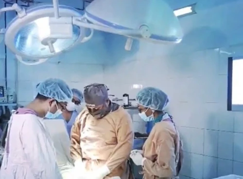
- Surgery is the primary focus of treatment for these stages. Methods range from essential removal to more intricate operations, such as neck dissection, contingent on the tumor's position and size. The objective is total extraction with clear margins. Surgical intervention achieves a high success rate in early-stage SCCHN, leading to a 5-year survival rate of around 70-90%.
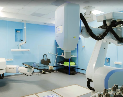
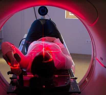
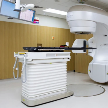
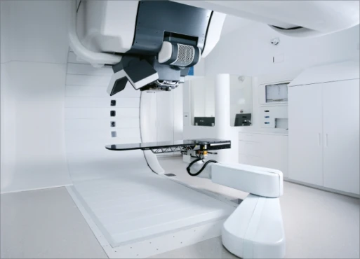
- Intensity-modulated radiation therapy (IMRT) is often employed after surgery or as the leading treatment option when surgery is not possible. IMRT accurately focuses on the tumor while protecting nearby healthy tissue, leading to reduced adverse effects. The effectiveness of radiation therapy in early-stage SCCHN is comparable to that of surgery, with high rates of local control achieved.
IMRT in a second-line treatment remains the preferred method.
Treatment protocol for advanced-stages SCCHN (III-IV stages)
First-line approach:
- Combined modality treatment: advanced-stage SCCHN typically necessitates a combination of surgical intervention, radiation therapy, and chemotherapy to achieve the best results. Generally, the 5-year survival rate for stage III and IV cancers ranges from 30% to 50%.
- Chemoradiation: concurrent chemoradiation is commonly used for patients who are not eligible for surgery. Combining chemotherapy with radiation therapy has demonstrated enhanced overall survival and control of the cancer in the local region.
- Chemotherapy: cisplatin is frequently used in combination with radiation as a chemotherapy treatment. It functions by causing damage to the DNA of cancer cells, ultimately resulting in their demise. The success rate for chemoradiation involving cisplatin is around 50-70% for advanced SCCHN.
Second-line therapy:
- Immunotherapy: immune checkpoint blockers like pembrolizumab and nivolumab have been authorized to treat recurring or spreading SCCHN. These medications function by inhibiting the PD-1/PD-L1 pathway, which cancer cells utilize to avoid detection by the immune system. In this scenario, the response rate to immunotherapy is approximately 20-30%, and some patients achieve long-lasting results.
Methods of Treatment for Specific Types of Tumors
Oropharyngeal Cancer
- First-line approach: Transoral robotic surgery in combination with radiation or chemoradiation. TORS enables the minimally invasive removal of tumors with reduced complications.
- Second-line therapy: Immunotherapy using checkpoint inhibitors for cases of recurring disease. Keytruda and Opdivo have demonstrated effectiveness in patients who have not responded to previous treatments.
Laryngeal Cancer
- First-line approach: Surgery that preserves organs, such as partial laryngectomy and radiation therapy. The goal is to maintain voice function while achieving control at a local level.
- Second-line therapy: Total laryngectomy for persistent or recurring disease. This procedure involves the complete removal of the larynx, requiring a permanent tracheostomy.
Nasopharyngeal Cancer
- First-line approach: Simultaneous chemoradiation, usually involving cisplatin. This combination is highly successful in treating nasopharyngeal cancer, resulting in high response rates and improved survival rates.
- Second-line therapy: Salvage surgery or additional cisplatin-based chemotherapy for cases that do not respond well to initial treatment.
Prognosis & Follow-up
How does cutting-edge science improve the lifespan and quality of life for those with the disease?
The prognosis for SCCHN differs greatly depending on the stage at diagnosis, where the tumor is located, and whether it is HPV-positive or negative:
- Early stages (I-II) have a 5-year survival rate of around 70-90%.
- In advanced stages (III-IV), the 5-year survival rate decreases to 30-50%.
- Oropharyngeal cancer with HPV-positivity generally shows better prospects compared to HPV-negative tumors, with a 5-year survival rate exceeding 80%.
Initial follow-up: patients with necessary physical exams and imaging are usually checked every 1-3 months during the initial year. Detecting recurrence early is essential for effective treatment.
Long-term follow-up: appointments every six months in the second and third years, then annually. Regular dental check-ups and nutritional assistance are vital to managing potential treatment complications.


