Disease Types & Epidemiology
How common is the disease?
Bone sarcomas (osteosarcomas) are rare tumors, accounting for less than 1% of malignant tumors. In the United States, approximately 62,038 individuals were estimated to be affected by bone and joint cancer in 2021 [SEER].
There are five types of bone sarcomas. However, it is crucial to know bone metastases (bone lesions resulting from the spread of cancer cells from other tumors, e.g., lung, prostate, breast, etc.) to a different part of the body are seen more frequently. Bone metastases are not bone sarcomas unless the primary tumor is a bone sarcoma.
Osteosarcoma is the most frequent type of primary tumor of bone. It is estimated that there are 2 to 3 new cases per million people every year; adolescents, particularly around the age of 15 to 19, are the most commonly affected age group.
Chondrosarcoma is the most frequently occurring bone sarcoma type of adulthood, with two new cases per million people diagnosed every year. The most common age at diagnosis is between 30 and 60.
Ewing sarcoma is the third most common bone sarcoma. It occurs more frequently in children and teenagers, usually diagnosed around 15 years of age, but it also occasionally appears in adults. It may involve any bone and soft tissues, but it is more common in the limbs (50%) and pelvic bones (25%); the ribs and vertebral column/spine may also be affected.
Giant cell tumor of the bone represents 5% of all primary bone tumors. It most commonly occurs between 21 and 30 years old and is more frequent in women.
Chordoma is a sporadic malignant bone tumor. It is diagnosed in one out of one million people every year. Common sites of origin are the sacrum (50%), the skull base (30%,) and the spine (20%). It is most frequently diagnosed in people at 60 years old. Skullbase lesions, however, generally affect a younger population, appearing around 50 years old in most cases, but it has also been reported in children.
Causes & Risk Factors
What is the primary issue of osteosarcoma?
Some risk factors for bone sarcoma have been identified, and the main ones are the following:
- Genetic predispositions: both inherited and acquired conditions may be associated with bone sarcoma.
- Li-Fraumeni syndrome: an inherited genetic condition caused by a mutation of a tumor suppressor gene (p53), which is a gene that helps to protect cells from becoming cancerous. Patients with this rare syndrome are more likely to develop several types of cancers, including bone sarcomas.
- Hereditary RB (retinoblastoma): a familial syndrome in which all body cells have a mutation in the RB1 gene. Patients usually develop malignant tumors of the retina (layer of nerve tissue in the back of the eye that receives images and sends them to the brain through nerves so images can be processed) in both eyes during infancy, and these children also have an increased risk of developing bone or soft tissue sarcomas, including osteosarcoma. A familial syndrome is a hereditary predisposition to a pattern of different tumor types and sites.
- Hereditary multiple exostoses (also known as multiple osteochondromatosis) is a rare inherited musculoskeletal disorder that causes short stature and deformities. In this condition, each osteochondroma has a minimal risk of developing into a bone sarcoma (most often a chondrosarcoma).
- Other rare inherited conditions, including Werner syndrome (hereditary disorder marked by rapid aging that starts in adolescence), Rothmund—Thomson syndrome (hereditary disorder that affects the skin, the bones, eyes, nose, hair, nails, teeth, testes, and ovaries) and Bloom syndrome (disorder marked by height that is shorter than average, a narrow face with redness and a rash, a high-pitched voice, and fertility problems), have also been linked to an increased risk of osteosarcoma.
- Osteosarcoma risk is higher in children and teenagers with Down's syndrome.
- Paget's disease of the bone is characterized by abnormal growth of new bone cells. The affected bones are fragile, misshapen, and more likely to break than normal healthy bones. Bone sarcomas (mostly osteosarcomas) develop in about 1% of people with Paget's disease, usually when many bones are affected. It primarily affects people older than 50.
- Ionizing radiation: exposure to ionizing radiation, such as X-rays and radiotherapy, can increase the risk of bone sarcomas even in the absence of other risk factors. Rarely, bone sarcomas can arise following exposure to radiation given to treat different cancers and often start in the area of the body that has been treated with radiation. The risk increases with the treatment dose and decreases with age. The average time between radiation exposure and diagnosis of bone sarcoma is about ten years. However, radiation exposure is an infrequent cause of bone sarcomas.
Clinical Manifestation & Symptoms
What signs should one anticipate while suspecting bone sarcoma?
Bone sarcomas might not cause symptoms for a long time, and inflammation (swelling and redness) will only be present if the tumor has progressed through the cortical bone. Symptoms depend on the size and location of the cancer. Bone pain is the most common symptom: it usually begins with a feeling of tenderness in the affected bone, which gradually progresses to a persistent ache. In some cases, the tumor can also weaken the bones, causing spontaneous fractures or fractures after a minor injury or fall. Nerve problems can be present due to constriction of the nerves by the tumor. Less common symptoms include fever, unexplained weight loss, fatigue/tiredness, or anemia (a reduction in red blood cells). Bone sarcomas may also be found by chance while investigating other symptoms or a routine operation.
Diagnostic Route & Screening
When, where, and how should bone sarcoma be detected?
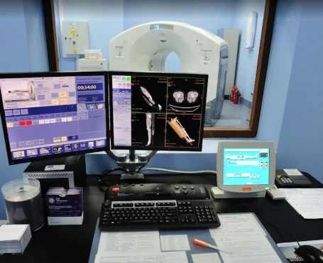
The diagnosis of bone sarcomas is based on the physical examination, radiological visualization techniques (bone X-rays / MRI / CT scan / PET scan / bone scintigraphy), histopathological examination (analyzing a piece of the tumor under a microscope), and blood tests (liver, kidney and blood cells function checking).
During physical examination, it's essential to evaluate the size and thickness of the swelling, its location and mobility, and the relation of swelling to the involved bone. Occasionally, this swelling may be painful or tender, but it may also be painless.
Choosing a visualization tool from a wide range of methods, it would be helpful to know the following information:
X-rays of the bone should always be the first test done since they can help to determine damage to the bones caused by cancer, new bone growth, or a bone fracture. Doctors can often recognize a bone tumor such as osteosarcoma (70%) based just on X-rays of the bone, but other imaging tests might also be needed.
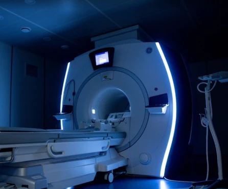
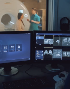
Magnetic Resonance Imaging (MRI) uses magnetic fields and radio waves to create a series of detailed pictures of the body's tissue. MRI of the affected bone, all other tissues surrounding it, and the adjacent joints is the best imaging test for diagnosing extremities (arms and legs) and pelvic tumors. It is an effective way of assessing the size and spread of cancer inside the bones or surrounding soft tissues.
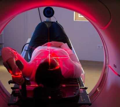
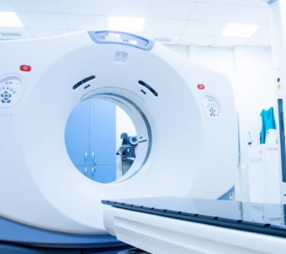
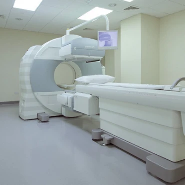
Computed Tomography scan (CT-scan) is an X-ray technique that produces detailed pictures of the inside of the body. You may be asked to drink a liquid called oral contrast and receive contrast liquid injected into your veins. This helps the organs or tissues show up more clearly and allows the visualization of calcifications (deposits of calcium) or bone destruction. CT scans can also be performed to check if bone sarcoma has spread to the lungs or other organs.
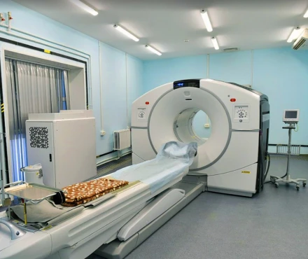
A Positron Emission Tomography (PET) scan is mainly used to determine if the sarcoma has spread to other body parts. It uses a substance that contains glucose, which is injected into the patient. This radiolabeled glucose-based substance is absorbed by cancerous cells, which are less able to eliminate it than normal tissues, so it remains "trapped" in cancerous tissues, making it visible. PET scans can also be used to examine the effect of the treatment on tumors so that cancer regression or progression is seen thanks to the radiolabeled glucose-based substance aforementioned.
Bone scintigraphy: a scan using a radiolabeled substance to determine whether other bones are affected. The radiolabeled substance travels to areas of bone changes, which appear brighter and indicate the tumor's possible spread.
During a biopsy for a histopathological examination, a small piece of the tumor might be taken from the lesion by two methods:
Fine needle / Core needle biopsy: cells of the tumor are removed using a needle. A local anesthetic is injected to numb the area before the biopsy, and several samples may be taken. The doctor may use imaging techniques such as ultrasonography or a CT scan to visualize and guide the needle into the right place if the tumor is located deeper within the body.
Incisional / Excisional biopsy: under anesthesia, surgical instruments are used to remove a piece of tissue from the tumor ("incisional") or the entire tumor ("excisional").
When a biopsy is taken through an incision, to be sure that the biopsy location is adequate and that the characteristics observed in that piece of tissue are likely to be similar to the characteristics of the whole tumor, it is recommended to make X-rays of the biopsy location and sometimes undertake another sample in case more material is required. In aggressive tumors, the biopsy track must be considered contaminated with tumors. It must be removed with the resection of the tumor sample to avoid local recurrences, including the possible channels through which drains have been placed. Biopsy tracks should be marked utilizing a small incision or ink tattoo to ensure the location can be recognized during the definitive procedure.
Staging and Treatment Approaches
What are the options for managing bone sarcoma?
Doctors use staging to assess the extension of the tumor in the body, which is an essential indicator of prognosis. The most widely used staging system for bone sarcomas is the TNM system. The combination of T (size of the tumor and invasion of nearby tissue), N (involvement of lymph nodes), and M (metastasis or spread of the tumor to other organs of the body) will classify it into one of the stages. For bone sarcomas, the TNM staging also considers the malignancy grade (G), a critical prognostic factor. Tumor burden and the presence of detectable distant disease are the two main factors considered in the clinical staging of these diseases.
The stage is fundamental to making the right decision about what treatment to use. The lower the stage, the better the prognosis. The tumor at:
Stage IA:
- is categorized as grade 1 or 2 (low-grade);
- is no more than 8 cm in its most significant dimension;
- has not spread to lymph nodes or other parts of the body.
Stage IB:
- is categorized as grade 1 or 2 (low-grade);
- is more than 8 cm in its most significant dimension or is located in different parts of the same bone;
- has not spread to lymph nodes or other parts of the body.
Stage IIA:
- grade 3 or 4 (high-grade);
- < 8 cm in its most significant dimension;
- has not spread to lymph ns or other parts of the body.
Stage IIB:
- grade 3 or 4 (high-grade);
- 8 cm in its most significant dimension;
- has not spread to lymph ns or other parts of the body.
Stage III
- grade 3 or 4 (high-grade);
- is located in different parts of the same bone;
- has not spread to lymph nodes or other parts of the body.
Stage IVA - has spread to the lungs.
Stage IVB - has spread to nearby lymph nodes or distant sites other than the lungs.
Phases of Treatment
How is bone sarcomas treatment structured?
Treatment plan for localized disease
Bone sarcomas are localized when confined to the primary site and have not spread to nearby tissues or other body areas. At this stage, the primary therapeutic goal is to remove the whole tumor by surgery whenever possible. Radiotherapy and chemotherapy can also be used to increase the chance of a definitive cure or reduce the risk that the tumor comes back.
Treatment for localized forms of bone sarcomas includes options that aim to act locally in the region affected by the disease.
Surgery
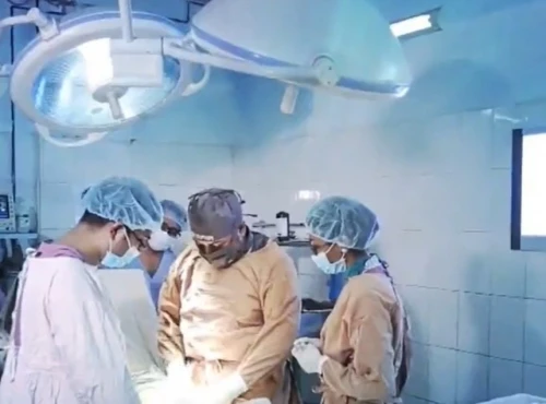
Most frequently, surgery is the standard treatment method used for localized bone sarcomas. As bone sarcomas are rare, surgery should be performed by a surgeon who specializes in treating this type of tumor. The goal of most bone sarcoma surgery is complete resection without leaving anything behind (microscopically negative margins), thereby reducing the risk of local recurrence. Today, it is rare to resort to amputations for limb bone sarcomas since currently, it is often possible to remove only the tumor and some of the surrounding tissue using a conservative approach, known as "limb-sparing" surgery, possibly with the contribution of other treatment modalities, including chemotherapy.
Several terms can define the completeness of the surgical resection:
- "R0" resection means the complete removal of all the tumors according to the analysis of the tissue margins done by a pathologist using a microscope;
- "R1" resection indicates that the margins of the resected parts show the presence of tumor cells when viewed microscopically;
- "R2" resection indicates a macroscopic residual disease (which means that a portion of the tumor visible to the naked eye could not be removed by surgery).
Small bone sarcomas can usually be effectively removed by surgery alone and curettage. Cryosurgery (using extreme cold to destroy abnormal tissue) can also be an option in selected cases. RI and R2 resection may need additional treatment by surgery, or another option is to treat the resected margin containing tumors with radiation and possibly chemotherapy.
Radiotherapy
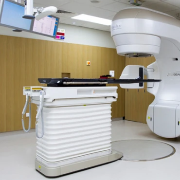
In bone sarcomas, radiotherapy may be used before (neo-adjuvant) surgery (to shrink the tumor and allow it to be eradicated) or after (adjuvant) surgery (to kill any remaining tumor cells); it may be considered in case of positive margins or case of residual macroscopic disease (when a portion of the tumor visible to the naked eye is still present). Radiotherapy may be performed in selected instances instead of surgery to achieve local disease control.
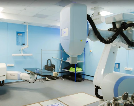
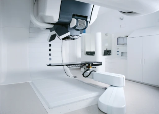
New radiation therapy techniques, like proton/ion beam radiotherapy, may be considered for some types of bone sarcomas. The difference between conventional and proton/ion beam radiotherapy is that high molecular weight particles, such as carbon ions and protons (hadrons), release almost all of their energy at the spot they are aimed at and not throughout their course as X-rays do. This causes less damage to surrounding healthy tissues.
Chemotherapy
Chemotherapy may be considered alone or combined with radiotherapy before or after surgery for localized disease. It is strongly recommended in these two situations:
- In osteosarcoma, chemotherapy has a well-established role in preventing local and distant relapses, and it is usually given both pre-operatively and post-operatively for a cumulative period of 6/10 months.
- In Ewing sarcoma, chemotherapy is usually given every three weeks, pre-operatively and post-operatively, for about 10/12 months with regimens including at least 5-6 different drugs. It may be used in combination with radiotherapy.
Chemotherapy is not routinely used in localized chondrosarcomas and is not an option in chordoma and giant cell tumors of the bone.
Treatment plan for advanced disease
Bone sarcomas are advanced when they have spread from where they started to other body parts. This is known as the metastatic phase. At this stage, the primary therapeutic goal is to control the disease, improving patients' quality of life by improving their symptoms.
Advanced disease is not treated exactly the same way every time in different patients. The best treatment strategy requires careful and individual consideration of the various options by a multidisciplinary team.
Occasionally, surgery may be considered in metastatic disease to relieve symptoms and may be curative in some cases, mainly when lung metastases are relatively few and slow-growing and are not accompanied by metastases located in organs other than the lungs.
Radiotherapy may also be given to relieve symptoms and control metastases, in particular bone metastases.
However, the primary treatment approach in the case of advanced disease is the use of systemic therapy, which includes both chemotherapy and molecularly targeted therapy (drugs that target specific proteins or cellular structures known to be involved in the growth and progression of cancer). Each type of drug works differently, altering how a tumor cell grows, divides, and repairs itself.
Chemotherapy
Chemotherapy is the mainstay of treatment for advanced disease, as the drugs administered enter the bloodstream and reach tumor cells throughout the body. The most commonly used chemotherapeutic drugs in bone sarcomas are doxorubicin and other anthracyclines, cisplatin, ifosfamide, cyclophosphamide, gemcitabine, docetaxel, etoposide, methotrexate, irinotecan, vincristine, and other vinca alkaloids.
Chemotherapy drugs can be given alone or in combination and may be provided as an outpatient or as an inpatient approach with admission to the hospital for a few days. Chemotherapy is given in cycles of treatment, and the chemotherapy regimen usually consists of several cycles over a set period: the number of cycles depends on the type, site, and size of bone sarcoma and how it responds to the drugs.
Targeted therapy
Targeted therapy may also be used to treat advanced disease. These therapies work by binding to a specific protein or cellular structure involved in tumor growth and progression, such as Denosumab for giant cell tumor bone treatment or pazopanib that targets multiple proteins involved in tumor growth and angiogenesis (the formation of new blood vessels that supply the tumor).
Prognosis & Follow-Up
How does cutting-edge science improve the lifespan and quality of life for those with the disease? How do you ensure continued health after treatment?
The 5-year relative survival rate in patients with bone sarcomas is 68,2% [SEER].
A proposed plan for follow-up care may involve assessment following the completion of chemotherapy, approximately every 2-3 months during the first two years and then every six months from years 3 to 5. Subsequently, evaluations can be scheduled every 6-12 months up to year 10, followed by intervals of 0.5-1-2 years based on local guidelines and individual considerations. If chest CT is used instead of chest X-rays, it should be performed using low-dose radiation techniques, especially in younger patients.
For individuals with low-grade bone cancer, the frequency of follow-up appointments may be reduced (e.g., every six months for two years and then annually). Late metastases, local recurrences, and functional impairments could manifest more than ten years after diagnosis; therefore, there isn't a universally accepted time to cease tumor monitoring [ESMO, 2021].


