Introduction & Classification of Chronic Myeloid Leukemia
Leukemia is a type of cancer of the blood. There are different forms of leukemia depending on what type of blood cell is affected. "Chronic" describes a gradual or slow progression, and "myeloid" denotes the origin of myeloid cells, immature cells that normally become mature red blood cells, white blood cells, or platelets. In chronic myeloid leukemia, the bone marrow produces too many myeloid blood cells, which are at various maturation stages, including cells known as immature granulocytes, metamyelocytes, and myeloblasts. Platelets and basophils (different myeloid cells responsible, in part, for the allergic response) are also often overproduced at diagnosis. Excess production of myeloid blood cells in the bone marrow ultimately prevents the normal production of red blood cells, essential in delivering oxygen to all cells in the body. It can also decrease the production of platelets or cause thrombocytopenia. Platelets play a critical role in stopping bleeding [ESMO, 2017].
The cause of chronic myeloid leukemia (CML) is known to result from a specific genetic abnormality that occurs in the blood stem cell. However, the exact cause leading to the cellular disbalance is not understood. The genetic abnormality is known as a translocation. For CML, specifically, genes from chromosome 9 are swapped with genes from chromosome 22. Translocation of the Abelson murine leukemia gene (ABL) on chromosome nine and the breakpoint cluster region (BCR) on chromosome 22, resulting in the Philadelphia chromosome [translocation of chromosomes 9 and 22, t(9;22)] can be detected in 95% of patients with CML either from cells circulating in the blood or the bone marrow.
The Philadelphia chromosome encodes a dysregulated tyrosine kinase (an enzyme in cells), which results in an abnormal behaviour of the cells affected. This includes the formation of immortalized cells, increased cell turnover and proliferation, and abnormal cell maturation.
Stages of CML
Unlike other cancers, which develop at a single site (such as breast cancer within the breast or prostate cancer within the prostate) and then spread (metastasize), malignant cells in patients with leukemia are considered to be present throughout the body at diagnosis due to their normal circulation in the bloodstream. For this reason, the prognosis is not determined by the extent of the spread of the disease. The stage of disease is determined by the "phase," including chronic, accelerated, and blastic phase or blast crisis. The majority of patients are diagnosed in the chronic phase. Patients are diagnosed with accelerated phase disease if the percentage of blasts increases to 15-29% in the blood or bone marrow, greater than 20% basophils develop in the blood, platelets either become severely elevated or low (but not as a result of therapy), or a clonal abnormality develops in addition to the Philadelphia chromosome. The most advanced stage of the disease is blast crisis, which is defined by an increase in bone marrow or peripheral blood blasts to at least 30%.
Algorithm of Diagnosis
What evaluations do CML patients undergo to identify the best treatment strategy?
Diagnosing CML involves a series of tests to confirm the disease, determine its stage, and identify the most appropriate treatment options [ashpublications.org].
- A thorough physical examination is performed to check for signs of lymphadenopathy (enlarged lymph nodes), hepatosplenomegaly (enlarged liver and spleen), and signs of anemia or thrombocytopenia.
- A complete blood count (CBC) and peripheral blood smear are essential to identify lymphocytosis and assess the overall health of blood cells. Flow cytometry is used to characterize the lymphocytes in a lab and confirm the diagnosis of CML by identifying specific cell surface markers (e.g., CD5, CD19, CD23, CD38).
- Although not always necessary for diagnosis, a bone marrow biopsy can help assess the extent of disease infiltration and the health of bone marrow function.
- Cytogenetic and Molecular Tests support the identification of genetic abnormalities that can influence prognosis and treatment decisions. Key genetic markers include:
- FISH (Fluorescence In Situ Hybridization) detects common chromosomal abnormalities such as del(13q), del(11q), and trisomy 12.
- PCR (Polymerase Chain Reaction) is used to identify specific mutations like TP53 and IGHV status.
- Next-Generation Sequencing (NGS) provides a comprehensive analysis of genetic mutations and helps identify targets for personalized therapy.
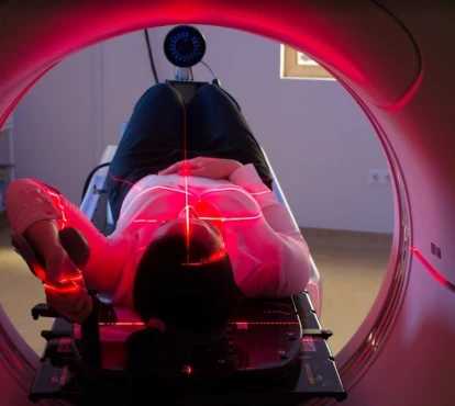
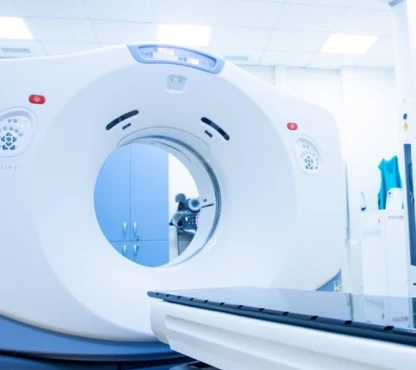
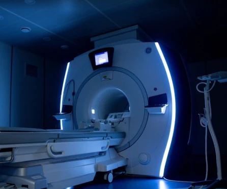
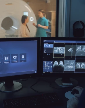
- Imaging Studies: CT scans, or ultrasounds may be used to evaluate the extent of lymphadenopathy and organ involvement.
Phases of Treatment
How is CML treatment structured?
Treatment for CML is tailored to the individual's disease stage, symptoms, and overall health [hematology.org].
Treatment for chronic phase CML
All CML patients should first be treated with a targeted inhibitor of the mutated tyrosine kinase, also known as the BCR-ABL tyrosine kinase. Imatinib is a first-generation oral tyrosine kinase inhibitor (TKI) that achieves an 8-year overall survival of nearly 90% of patients. Second-generation tyrosine kinase inhibitors, such as dasatinib or nilotinib, may also be considered for all patients with CML upon diagnosis. More specific TKI include ibrutinib (acalabrutinib - as an alternative) for CML patients with del(17p) or TP53 mutations,
Other agents, including interferon and hydroxyurea, have a limited role in first-line therapy. Hydroxyurea is used to rapidly lower disease burden and white blood cell counts.
Response assessment is based on three levels of response: hematologic response, cytogenetic
response and molecular response, as described below:
1. Hematologic response:
- The peripheral WBC level and platelet count are monitored every two weeks after the initiation of therapy.
- A complete hematologic response (CHR) – meaning normalization of blood environment - is the association of
- a total WBC <10x10<sup>9</sup>/l,
- a WBC formula with no present immature granulocytes and <5% of basophils,
- platelets count <450x10<sup>9</sup>/l,
- and non-palpable spleen.
2. Cytogenetic response (CgR):
- Cytogenetics is monitored after three months, six months, 12 months, and 18 months of therapy with a TKI. Until a complete cytogenetic response (CCgR) is achieved, cytogenetics should be repeated at least every six months.
- A complete cytogenetic response (CCgR)is defined by the inability to detect the Philadelphia chromosome by analyzing the chromosomes metaphases in a lab.
- A partial cytogenetic response (PCgR) is defined as the presence of 1%–35% positive Philadelphia chromosome metaphases.
3. Molecular response
- Monitoring of molecular response is done with PCR, which quantifies the BCR-ABL/ ABL mutations.
- It should be repeated at least every three months until a significant molecular remission is achieved.
- Major molecular remission is defined by a PCR result with BCR-ABL/ABL <0.10% on the International Scale.
Once a complete cytogenetic response and a significant molecular remission have been achieved, cytogenetic monitoring should be performed every 12 months, and molecular tracking by PCR should be performed every six months.
Treatment for accelerated phase or blast crisis CML
Treatment with a tyrosine kinase inhibitor can be initiated in patients who have not already been treated with a tyrosine kinase inhibitor. Change to another tyrosine kinase inhibitor, or chemotherapy can be considered for patients already treated with a tyrosine kinase inhibitor. However, those options are effective for only a limited time. Allogeneic bone marrow transplant is the only established curative therapy for CML in either of these phases of disease.
Treatment of patients who cannot tolerate tyrosine kinase inhibitors
Patients who develop severe side effects from first-generation tyrosine kinase inhibitors, such as severe rash, severe edema (swelling of the legs), or fluid accumulation in the lungs, should be first treated with second-generation tyrosine kinase inhibitors. Most patients who cannot tolerate first-generation tyrosine kinase inhibitors can be successfully treated with second-generation tyrosine kinase inhibitors without side effects. The dose of second-generation tyrosine kinase inhibitors can be reduced without decreasing efficacy to reduce the risk of side effects. For patients who cannot tolerate three tyrosine kinase inhibitors, a new therapy, omacetaxine, was recently shown to be effective and tolerable. In rare cases, patients who cannot tolerate all tyrosine kinase inhibitors should be considered for a bone marrow transplant from a sibling or unrelated donor.
Prognosis and Survival Rates
What are the survival rates and factors affecting prognosis?
The prognosis for CML varies widely based on genetic factors, disease stage, and response to treatment. The overall five-year survival rate for CML is approximately 85%, but this can vary significantly:
- In patients with favourable cytogenetics (e.g., del(13q) only), there is a more prolonged median survival, often exceeding ten years after the treatment ends.
- Patients with high-risk cytogenetics (e.g., del(17p) or TP53 mutations) - shorter median survival, often 2-5 years without effective targeted therapy [cancer.org].
Untreated patients with chronic phase CML will progress to the accelerated phase in 3-5 years. Patients
diagnosed with accelerated phase have a median survival of 4 to 6 months without treatment. Survival is further limited if a blast crisis occurs, with a median survival among untreated patients of 2 to 4 months.
A patient's prognosis is best predicted by the patient's characteristics (including percentage of basophils as well as spleen size). Multiple scoring systems using patients and disease characteristics have been developed, which estimate the likelihood of response to therapy and survival. When using the most up-to-date EUTOS risk score, five-year, progression-free survival was significantly better in the low than in the high-risk group (90% vs. 82%), but overall survival was only slightly lower than that of the standard, healthy population [European Leukemia Net, 2024].
Follow-Up and Monitoring
How do you ensure continued health after treatment?
In CML patients, the follow-up recommendation includes physical examination and CBC every three months for five years and repeated bone marrow biopsy with PCR cytogenetic testing only in case of treatment failure or in case of unexplained thrombocytopenia every three to six months until the complete (normal) cytogenetic response is achieved with no T315I BCR-ABL mutation detected [American Journal of Hematology, 2022].


