Introduction & Classification of Follicular Lymphoma
Follicular lymphoma develops in the white blood cells, lymphatic system, and bone marrow. Follicular lymphoma is a well-defined subtype (low grade) of Non-Hodgkin Lymphoma (NHL), with cells of the lymphoid tissues in the lymphatic system multiplying uncontrollably to cause tumors to grow eventually.
Different types of lymphoma can develop from each type of lymphocyte, but follicular lymphoma arises in particular from B-lymphocytes. The malignant cells in lymphoma grow in clusters to form nodules. Some organs are also part of the lymphatic system and are partially constructed by lymphoid tissue, which includes the spleen, thymus, tonsils, and adenoids. The lymphatic system filters blood lymph (the liquid circulating in lymph vessels), drains fluid from tissues to the bloodstream, and fights infections. Since lymphoid tissue is found throughout the body, follicular lymphoma can begin in almost any body part. The bone marrow may become invaded by lymphocytes that do not function properly. As the bone marrow also produces platelets to stop bleeding critically and red blood cells to deliver oxygen to all cells in the body, excess accumulation of lymphocytes prevents the normal production of red blood cells and platelets. Follicular lymphoma is usually slow-growing.
Compared to breast cancer in women or prostate cancer in men, NHLs are not common. Nevertheless, they are the sixth most common cancer in Europe. They account for around 3% of all cancers; follicular lymphomas represent approximately 25% of all NHLs. In Western Europe, follicular lymphoma is the second most frequent subtype.
The number of patients diagnosed with follicular lymphoma yearly has increased from 2-3 cases per 100,000 people in the 1950s to 5-7 cases per 100,000 people.
In general, the risk of getting NHL increases with age. There is a 5-7-fold increase in cases among patients older than 65 years.
Follicular lymphoma staging is the process of determining whether the tumor has spread and, if so, how far. It is essential to know the stage of the disease to plan the treatment. The staging system used to describe the spread of follicular lymphoma is called the Ann Arbor Staging System. It uses Roman numerals (I-IV) for different stages:
- Stage I - The lymphoma is in one group of the body's lymph nodes, such as the groin or neck, or one organ of the lymph system.
- Stage II - Two or more groups of lymph nodes, or one organ close to the affected lymph nodes, and one or more groups of lymph nodes on the same side of the diaphragm contain lymphoma cells. The diaphragm is the muscle that divides the chest and abdomen. For example, lymphoma might be above the diaphragm in lymph nodes in the neck and armpits. Or, the lymphoma might be below the diaphragm in the lymph nodes in the groin and abdomen.
- Stage III - The lymphoma is in lymph nodes on both sides of the diaphragm. It may have also spread into an organ close to the affected lymph nodes or spleen.
- Stage IV - The lymphoma is in stage IV if the lymphoma involves the bone marrow or distant organs.
Follicular lymphoma stages are also noted by the presence or absence of certain findings and/or symptoms of the disease:
- A lymphoma that affects organs or tissues other than the lymph nodes has an "E," for
- extranodal, added to its staging nomenclature.
- If a nodal mass is at least 7.5 cm in diameter, it translates as a bulky disease.
- If it affects the spleen, an "S" is added.
- If the patient has a fever, night sweats, or unexplained weight loss, the letter "B" is added.
- If none of these is present, an "A" is added.
The grading from the WHO organization could be 1, 2, 3A, and 3B to reflect the number of lymphoma cells or blasts under the microscope using maximal magnification. Grade 3B is the highest grade and is considered an aggressive lymphoma.
A Follicular Lymphoma-specific International Prognostic Index (FLIPI) should be determined for predictive purposes. FLIPI allows us to identify the disease's progression risk after treatment and adapt treatment and follow-up accordingly. The parameters used in the original version of FLIPI 1 are > four involved nodal sites, age >60 years, elevated lactate dehydrogenase (LDH) level, stage III or IV disease, and hemoglobin <12 g/dl. Each of the above characteristics is assigned one point, ranging this index score between 0 and 5. For each index relevance, the 0-1 risk is considered low, the two risks as intermediate, and the 3-5 risk as high.
Pediatric follicular lymphoma is characterized by a localized disease that is histologically more aggressive, and some have specific molecular features. However, pediatric follicular lymphoma shows a much more indolent course and should be managed with local therapy only.
Phases of Treatment
How is follicular lymphoma treatment structured?
Treatment plan for stage I-II follicular lymphoma
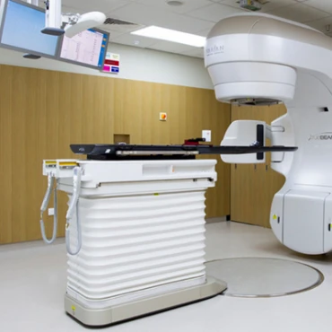
In the small proportion of patients with limited non-bulky stage I-II disease, the administration of radiotherapy targeting the site of the involved lymph nodes has curative potential.
In selected cases, watchful waiting may be discussed to avoid the side effects of radiation and it could be of the same efficacy as active treatment.
The presence of bulky, large tumors or with two or more sites involved helps identify patients who could benefit from chemotherapy (vincristine, doxorubicin) and monoclonal antibody rituximab. In this case, the role of radiotherapy may be considered after this initial treatment if the sites of involved lymph nodes are located so that radiotherapy can be given without significant side effects.
Treatment plan for stage III-IV follicular lymphoma
The disease could disappear or regress without treatment in 10-20% of lymphoma cases. Early initiation of therapy in asymptomatic patients did not show any improvement in survival. Therefore, watchful waiting is recommended.
Treatment should only be utilized due to symptoms, including B-symptoms (fever for an unknown reason, drenching night sweats, and undesired or unintentional weight loss), impairment in blood cell formation, bulky disease, compression of important organs, abnormal presence of fluid in the abdominal cavity (ascites), or in the space between the lungs and thoracic wall (pleural effusion) and rapid progression of the disease.
The induction treatment (chemo with vincristine, cyclophosphamide, and prednisone) is the first step in reducing the number of cancer cells. From there, a consolidation phase (with rituximab) further reduces the number of cancer cells and increases the probability of the lymphoma not returning. This is followed by a maintenance phase with goals to maintain the remission and prevent a relapse.
Relapsed or Refractory Follicular lymphoma treatment approach
Relapse is the reappearance of the disease. A repeated biopsy is strongly recommended to know if the lymphoma that relapsed turned into an aggressive form.
The treatment given when the disease relapsed is called salvage treatment, and selection depends on the effectiveness of the previous treatments administered. In early relapse (< 12- 24 months disease-free), the disease might be resistant to the drugs used previously. As a result, a regimen of different drugs to overcome resistance is preferred. An example comes from bendamustine after CHOP (cyclophosphamide, doxorubicin, vincristine, and prednisone) and vice versa. Rituximab could be used again for the patient if it had previously achieved a disease-free period of more than 6-12 months.
Radioimmunotherapy (radioactive substance combined/attached to a monoclonal antibody, such as <sup>90</sup>Y-Ibritumomab-tiuxetan (Zevalin) and <sup>131</sup>I-tositumomab (Bexxar)) represents an effective approach, especially in patients over 65 years of age with other diseases present who are not eligible for chemotherapy.
Rituximab maintenance for up to 2 years as a single treatment every three months can be administered in patients who received it as a part of induction and did not receive it in the first-line treatment.
In young patients, high-dose chemotherapy with a stem cell transplantation using the patient's stem cells could be considered. Research indicates that treatment combination for young cancer patients delays the progression of the disease and prolongs survival. However, it is not always required now. Instead, rituximab is widely used for patients, especially in patients experiencing late relapses.
Prognosis, Survival Rates, and Follow-up
What are the survival rates and factors affecting prognosis?
Generally, for people with follicular lymphoma, around 85% of people survive their cancer for five years or more after diagnosis. They will be from the low-risk group (100%), intermediate-risk group (90%), and high-risk group (75%).
Patients should undergo:
- History-taking, monitoring of symptoms, and physical examination every three months for two years, every four to six months for an additional three years, and subsequently once a year.
- Blood count and other routine blood analyses are performed every six months for two years and only if suspicious symptoms appear.
- Evaluation of thyroid function at one, two, and five years if the patient received irradiation to the neck.
- Radiological and ultrasound studies are recommended every six months for two years and annually thereafter. However, CT scans are not mandatory outside clinical trials.


