Disease Types & Epidemiology
How common is the disease?
Multiple myeloma (MM) is a form of cancer originating from plasma cells, a type of blood cell located in the bone marrow. These cells play a role in producing antibodies to combat infections. However, in myeloma cases, cells turn cancerous and grow out of control. This type of cancer represents around 1.8% of all malignancies and 10% of hematologic tumors, making more than 35 700 new cases each year in the United States. Multiple myeloma has an average five-year survival rate of 61.1%, according to the National Cancer Institute, and an estimated prevalence of 179,000+ people living with myeloma in the US. It stands as the prevalent blood cancer following non-Hodgkin lymphoma. Typically, this disease impacts individuals with an average age, at diagnosis being 69 years old. Men and African Americans tend to have incidence rates compared to Asians, who exhibit rates [SEER].
Causes & Risk Factors
What is the primary issue of myeloma?
To date, no cause for myeloma has been identified. Research suggests that the disease could be related to a decline in the immune system, certain occupations, exposure to certain chemicals, and radiation exposure; these connections are not strong. In most cases, multiple myeloma develops in individuals with known risk factors. Multiple myeloma may be the result of several factors acting together. It is uncommon for myeloma to develop in more than one family member.
Most multiple myelomas arise from a benign condition known as Monoclonal Gammopathy of Uncertain Significance (MGUS). People affected by this condition have abnormal monoclonal protein production without any symptoms. The majority of people with this condition will never develop symptomatic multiple myeloma. In most cases, MGUS is discovered by accident during routine blood tests [ESMO, 2021].
Clinical Manifestation & Symptoms
What signs should one anticipate while suspecting myeloma?
If MGUS progresses and develops into multiple myeloma, prompt treatment can prevent the development of disease symptoms. Two groups of specific clinical signs accompany multiple myeloma [Cancer.net]. These groups’ sources are connected to the exact pathological segment of the MM, such as:
- Symptoms caused by bone marrow infiltration:
- Fatigue: this is the physical feeling of being tired even after rest. It is related to anemia (low hemoglobin level) and the abnormal presence of multiple myeloma in the body.
- Bone pain and fractures: sometimes progressively intense bone pain is present, and it rarely responds to common painkillers. This pain is often felt in the spine, ribs, or hip bones and could result from bone fractures.
- Infections: infections may occur more frequently, and these infections may take longer to heal than in the past in the same person. This is related to both a decreased white cell count and the abnormal function of plasma cells.
- Bleeding: rarely, abnormal bleeding may occur (for example, during teeth brushing) or notification about easily occurring bruising or hematomas. These events are related to low platelet count and to abnormalities in mechanisms responsible for stopping the bleeding because of monoclonal protein in the blood.
- Symptoms or signs related to excessive monoclonal protein production:
- Mild to severe kidney problems: this condition is caused by direct damage from monoclonal protein filtered by the kidneys. Usually, this condition does not cause symptoms until the damage is severe.
- Amyloidosis: this is caused by abnormal accumulation of monoclonal protein in specific body sites (heart, kidney, etc.). The abnormal stores of the protein can cause chronic inflammation and organ damage.
- Peripheral neuropathy: this results from nerve damage caused by monoclonal protein. Sensory disturbances (tingling, altered heat perception in hands and feet, etc.) are the most common symptoms.
Diagnostic Route & Screening
When, where, and how should myeloma be detected?
The diagnosis of multiple myeloma is based on:
- the detection of monoclonal protein in the blood or 24-hour urine samples (obtained by a test called protein electrophoresis with subsequent measurement of the levels of serum free light chain with immunofixation to identify the type of monoclonal protein present);
- the percentage of myeloma cells in the bone marrow is analyzed using a bone marrow aspirate and/or biopsy.
Both procedures are minimally invasive and last for about 10-15 minutes. Local anesthesia is used before the procedure; a mild burning sensation should be expected. The samples obtained are necessary to quantify the percentage of plasma cells in the bone marrow and perform genetic tests, such as Fluorescence In situ Hybridization (FISH). These tests are helpful as they provide additional information on the prognosis of the disease, which is vital since they could influence the choice of treatment.
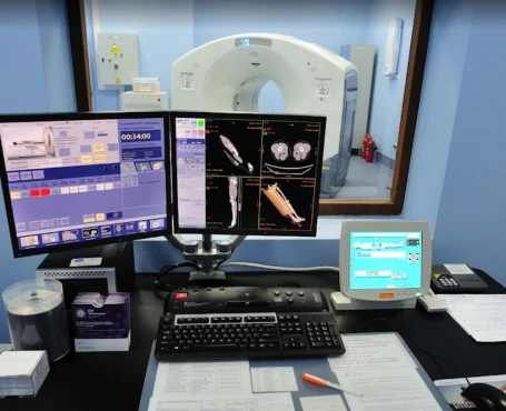
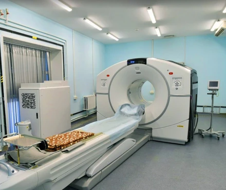
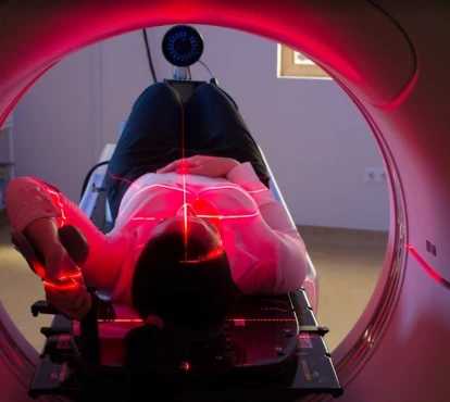
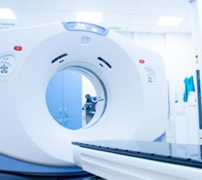
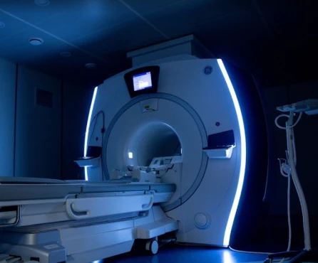
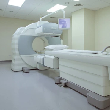
- Evaluation of bone lesions: a complete radiological skeletal bone scan is necessary to identify possible fractures or areas of disease infiltration. Magnetic resonance imaging (MRI) of the spine and pelvis is more sensitive than X-ray in detecting bone lesions. This helps identify lesions when they are not yet causing symptoms. A whole-body low-dose CT or PET scan may also be needed to evaluate bone lesions.
- Blood tests: complete blood cell count, calcium, creatinine, albumin, and beta-2-microglobulin levels are all necessary to examine if the disease is symptomatic and for prognostic reasons.
These blood tests allow differentiation between three conditions:
4.1) Monoclonal Gammopathy of Uncertain Significance (MGUS): a benign condition that rarely develops into multiple myeloma and is characterized by serum monoclonal protein <3g/dl; tumoural bone marrow plasma cells <10%; normal calcium levels, normal kidney function, normal hemoglobin levels, and no bone lesions.
4.2) Asymptomatic (smoldering) multiple myeloma: a pathologic condition that progresses to
multiple myeloma at a rate of 10% per year over the first five years following diagnosis. It is characterized by serum monoclonal protein >3g/dL or urinary monoclonal protein >500mg/24 hour and/or tumoural bone marrow plasma cells 10-60% without any multiple myeloma defining events (listed in the table below) or amyloidosis.
4.3) Multiple myeloma: the symptomatic condition which requires treatment. It has the same
features of asymptomatic (smoldering) multiple myeloma plus multiple myeloma defining
events, such as:
- Hypercalcemia - serum calcium >1mg/dL higher than the upper limit of normal or >11mg/dL;
- Kidney function decrease - creatinine clearance <40mL per min or serum creatinine >2mg/dL;
- Bone lesions on skeletal X-ray, CT, PET-CT or MRI scan;
- Bone marrow plasma cells excess >60%;
- Very high serum-free light chain ratio >100.
- Cytogenetic lab test - Fluorescence in Situ Hybridization (FISH) analysis - highlights the chromosomes in the biopsy sample. This makes it possible to examine them in sufficient detail to identify the nature of any chromosomal abnormalities (CA), which can include chromosomal translocations (when a piece of one chromosome swaps places with a piece of another chromosome), chromosomal deletions (when a piece of a chromosome is missing), and an increase in the number of chromosomes (also called hyperdiploidy). The high-risk CA markers in multiple myeloma are deletion 17p, translocation (4;14) and translocation (14;16) [cancer.gov].
Staging and Treatment Approaches
What are the options for managing myeloma?
Treatment for multiple myeloma is not necessary when there are no symptoms. Moreover, disease staging and cytogenetics are unnecessary for asymptomatic (smoldering) multiple myeloma. Thus, information about the stage of the disease is necessary when multiple myeloma is symptomatic, and treatment has to be started.
Information about the stages is essential to selecting the proper treatment. The lower the stage, the better the prognosis. The Revised International Staging System (ISS) is a beneficial score for this disease. It relies only on the serum levels of albumin and beta-2-microglobulin.
Stage I - Serum beta-2-microglobulin <3.5 mg/dl + serum albumin > 3.5 g/dl and standard risk CA by iFSH and normal lactate dehydrogenase (LDH).
Stage II - Serum beta-2-microglobulin 3.5 – 5.5 mg/dl and serum albumin > 3.5 g/dl.
Stage III - Serum beta-2-microglobulin > 5.5 mg/l and either high-risk CA or high LDH.
Phases of Treatment
How is MM treatment structured?
First-line treatment plan for autologous stem cell transplantation candidates
Patients in good physical condition (or those younger than 65) who are candidates for autologous stem cell transplantation usually receive an induction treatment. The aim of this is to reduce the disease burden before the transplantation. Once the disease burden is reduced, the goal is to maintain a response for as long as possible with an autologous transplant.
Induction treatment is usually composed of a three-drug regimen, including combinations of Bortezomib, thalidomide, Lenalidomide, cyclophosphamide, and dexamethasone.
One treatment cycle usually lasts 21 or 28 days. Response to treatment is assessed before each cycle. The total number of cycles required to complete the induction treatment ranges from 4 to 6, depending on the type of response, therapy, and patient’s health status.
Therapy efficacy is measured by the reduction of monoclonal protein, measured in blood serum or urine:
- Stringent complete response: disappearance of monoclonal protein in serum or urine (immunofixation negative, average free light chain ratio, absence of tumor plasma cells in the bone marrow);
- Complete response: disappearance of monoclonal protein in serum and/or urine (immunofixation negative, abnormal free light chain ratio, <5% plasma cells in bone marrow);
- Excellent partial response: 90% or greater reduction in serum protein plus urine protein <100mg per 24 h or serum and/or urine protein detectable by immunofixation but not with electrophoresis;
- Partial response:
-- more than 50% reduction of serum protein and reduction in 24 h urinary protein by >90% or to <200mg per 24 h;
-- in patients without measurable serum and urine monoclonal protein levels, the difference between involved and uninvolved free light chain levels (FISH test) can be used;
-- In patients without measurable serum and urine monoclonal protein levels and without measurable involved free light chain levels, bone marrow plasma-cell percentage can be used.
- Minimal response: the same as partial remission, but serum or urine protein reduction comprised 25% - 49%.
- Stable disease: response criteria not fulfilling the definition of complete response, perfect partial response, partial response, minor response.
- Progressive disease: any one or more of the following criteria:
- Increase of 25% from the lowest confirmed response value in one or more of the following criteria: serum monoclonal protein or urine monoclonal protein;
- In patients without measurable serum and urine monoclonal protein levels, the difference between involved and uninvolved free light chain levels can be used;
- In patients without measurable serum and urine monoclonal protein levels and without measurable involved free light chain levels, bone marrow plasma-cell percentage can be used;
- Appearance of a new bone lesion(s) or increase of existing lesion(s) if this is the only measure of disease;
- Increase in circulating plasma cells if this is the only measure of disease.
After the induction therapy, a consolidation phase is necessary to prolong the interval that patients remain free from disease. In multiple myeloma, consolidation is obtained with autologous stem cell transplantation. This process is preceded by collecting autologous (of the patient) stem cells by a procedure called apheresis. To stimulate the release of stem cells from the bone marrow to the bloodstream, the patient receives a growth factor (granulocyte-colony stimulating factor, G-CSF) alone or in combination with chemotherapy (cyclophosphamide). After a few days, when the number of stem cells rises, the patient receives the apheresis procedure. The number of stem cells can be determined by blood tests. Peripheral blood is filtered, and stem cells are collected and frozen. Once the collection has been done and the patient has recovered from the procedure, they can be admitted for autologous transplantation. This procedure consists of administering high-dose chemotherapy (usually with a drug called melphalan) followed by reinfusion of the patient’s own stem cells.
If the first transplant does not give a complete or almost complete response, a second autologous transplantation can be performed usually within 3-6 months after the first.
Allogeneic stem cell transplantation (from a donor) should only be carried out in the context of a clinical trial.
First-line treatment plan for non-transplant candidates (70 years and older or patients in poor physical condition)
The initial treatment is a three-drug regimen (bortezomib or thalidomide, melphalan, prednisone) or two-drug regimen (any pair of lenalidomide, dexamethasone, bendamustine, melphalan, prednisone) for frail patients.
Second-line treatment plan for relapsed or refractory disease
There are more advanced drugs with immunomodulatory effects (pomalidomide), chemotherapeutics (doxorubicin), or proteasome inhibitors (carfilzomib blocks protein synthesis in cancer cells).
Autologous stem cell transplantation could also be used in selected cases (in those with good response to previous autologous transplants and disease response of longer than two years).
Treatment of multiple myeloma complications
Impaired kidney function: almost fifty percent of patients affected by multiple myeloma have impaired kidney function. Treatment may vary depending on the grade of kidney impairment. Along with systemic therapy, oral and intravenous hydration or even dialysis could be part of the treatment. It is fundamental to avoid the use of non-steroidal anti-inflammatory drugs, such as aspirin or nimesulide, as they may cause kidney damage.
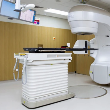
Bone pain or bone lesions: bone damage is frequent in multiple myeloma. It can be asymptomatic, or it may cause pain. In some cases, bone fractures can be the initial manifestation of multiple myeloma, and in this case, an orthopedic intervention is necessary. Besides surgical intervention, radiotherapy can also be helpful.
If bone lesions are absent but there are early signs of bone erosion, therapy with bone-strengthening drugs is suggested. Bisphosphonates are the primary drugs used for this purpose. Zoledronate or pamidronate is given through intravenous infusion. This treatment should last two years, and jaw infections should be excluded before starting these drugs.
Increased blood calcium level: this is due to bone erosion. The extent of the increase can vary. Intravenous fluids and bisphosphonates are required in case of very high calcium levels.
Anaemia: this is related to low red blood cell count. There are several causes of anemia in multiple myeloma. Bone marrow infiltration by abnormal plasma cells and/or kidney damage are the most frequent causes. Blood transfusions can be necessary in very severe cases. Administration of erythropoietin, a drug that stimulates red blood cell production, may decrease the need for transfusions.
Infections: both chemotherapy and multiple myeloma can weaken the immune system. For this reason, anti-infective drugs may be administered to prevent infection. The influenza vaccine helps decrease the rate of respiratory illness.
Spinal cord compression: the cause of this complication is the presence of a localized mass (plasmacytoma) at the backbone level, which compresses the spine. Backbone fractures can also cause this. Symptoms are localized pain or nervous symptoms such as leg tingling or muscular weakness. The patient should seek medical attention immediately if they experience these symptoms, as this complication can lead to irreversible paralysis if not treated. Corticosteroids, radiotherapy, or even surgery are therapies available to treat this condition.
Prognosis & Follow-Up
How does cutting-edge science improve the lifespan and quality of life for those with the disease? How do you ensure continued health after treatment?
5-year survival rates for myeloma based on the SEER stage show 79% for localized myeloma, 57% for distant MM, and a combined rate of 58% for all stages [SEER].
The majority of patients undergoing treatment for systemic myeloma also receive regular intravenous bisphosphonates. However, the long-term use of these drugs may lead to kidney problems or jaw osteonecrosis in a small number of patients, potentially impacting future use.
Follow-up care typically involves blood tests, periodic imaging scans, and bone marrow evaluation every 1 to 3 months. Given the long-term nature of myeloma treatment, ongoing monitoring is essential to detect any recurrence.


