Introduction & Classification of Acute Myeloid Leukemia
Acute Myeloid Leukemia (AML) is a type of blood cancer that starts in the bone marrow—the soft inner part of bones where new blood cells are made. AML progresses rapidly and can affect different types of blood cells. It is most common in adults, with a median age of diagnosis around 68 years, but it can also occur in children and adolescents. In the United States, AML accounts for approximately 1% of all cancer cases and about 32% of all leukemias. The American Cancer Society estimates that in 2023, there will be about 20,380 new cases of AML and about 11,540 deaths due to AML in the United States alone [cancer.org].
AML occurs slightly more frequently in men than women and is more common in individuals with a history of certain blood disorders, such as myelodysplastic syndromes (MDS), polycythemia vera, and essential thrombocythemia, previous chemotherapy or radiation therapy, or exposure to certain chemicals like benzene.
AML is classified into several subtypes based on the type of cell from which the leukemia develops and specific genetic markers. Here are the main subtypes:
- AML with recurrent genetic abnormalities includes AML with translocations - the movement of two chromosomes next to each other - such as t(8;21) and inversions – the cross-changes of chromosome pieces – like inv(16), which generally have a better prognosis. Acute promyelocytic leukemia (APL) subtype contains the t(15;17) translocation and is associated with a unique treatment approach, including vitamin therapy, which causes the leukemia cells to mature and has high cure rates.
- AML with myelodysplasia-related changes characterized by myelodysplasia – when white blood cells are abnormal in their shape and size - occurs in patients with a history of myelodysplastic syndrome (MDS) and typically has a poorer prognosis. Genetic mutations like those in the TP53 gene are common in this subtype.
- Therapy-related AML develops after treatment with chemotherapy or radiation for other cancers and has a generally poor prognosis. Mutations in the RUNX1 gene are frequently observed in this group as a hallmark of the AML subtype.
- AML not otherwise specified: This includes several subtypes classified by the appearance of the leukemia cells under the microscope. Among other genetic mutation markers in this AML group, NPM1 and FLT3 genetic abnormalities are crucial in determining prognosis and treatment options.
Stages of AML
While AML does not have formal stages like solid tumors, it is categorized based on the phases of treatment and the disease's extent at diagnosis:
- Stage I: AML confined to the bone marrow and blood.
- Stage II: AML with involvement of extramedullary sites such as the central nervous system.
- Stage III: Extensive involvement of organs such as the liver and spleen.
- Stage IV: AML with the widespread participation of multiple organs and tissues.
Algorithm of Diagnosis
What evaluations do AML patients undergo to identify the best treatment strategy?
Diagnosing AML involves several steps to confirm the disease and determine the next therapeutic steps [hematology.org]:
- Clinical Examination to check for signs of anemia, infections, and bleeding. The doctor may also look for enlarged lymph nodes, liver, or spleen.
- A complete blood count (CBC) measures the levels of different blood cells. Occasionally, a patient may have a complete blood count done for another reason, which will be the first indication of a possible AML based on laboratory findings alone. In addition to identifying a low red blood cell count or platelet count, the complete blood count may detect leukemia cells circulating in the blood as part of the white blood cell count. Immature white blood cells increasing at an abnormal rate are more significant than the more mature normal white blood cells found in circulation.
- Blood coagulation tests must be performed for patients with acute promyelocytic leukemia (APL) since coagulation disorders are widespread in this type of AML. Such tests must be performed before insertion of central intravenous lines.
- A bone marrow biopsy is performed if a diagnosis of AML is suspected based on symptoms and the white blood cell count. A bone marrow biopsy is a minimally uncomfortable procedure lasting about fifteen minutes. Local anesthesia is used for the procedure, and sharp pain is usually not experienced. The pathologist in a lab can determine what type of AML a patient has and further identify the genetic abnormalities of the leukemia by looking closely at the chromosomes through a microscope and molecular tests.
- Cytogenetic and Molecular Tests analyze the chromosomes of the leukemia cells to identify specific genetic abnormalities. For example, the t(8;21) and inv(16) translocations are common in particular subtypes of AML. Mutations in genes such as FLT3, NPM1, and CEBPA are also critical for prognosis and treatment planning. Specific tests include karyotyping (chromosome visualization), fluorescence in situ hybridization (FISH) (fluorescence staining of DNA), Polymerase Chain Reaction (PCR) (target AML mutations detection, such as FLT3-ITD mutation or the NPM1 mutation), Next-Generation Sequencing (NGS) (precise genetic method to diagnose of all sorts of gene mutations).
- Lumbar Puncture checks for the presence of leukemia cells in the cerebrospinal fluid, indicating central nervous system involvement.
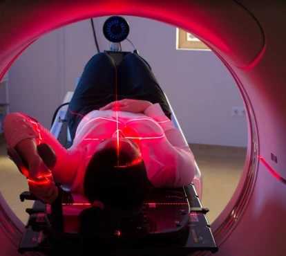
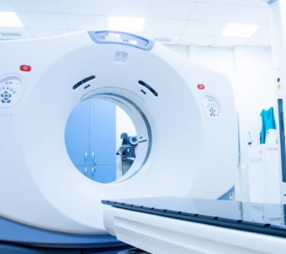
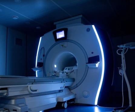
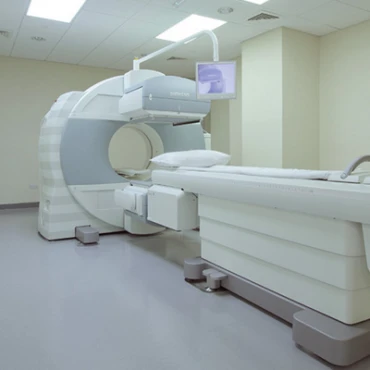
- Imaging Studies, such as CT scans or MRIs, may be used to detect any enlarged organs or lymph nodes and evaluate the disease's spread.
Phases of Treatment
How is AML treatment structured?
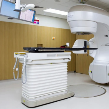
Treatment of AML is tailored to the individual based on the diagnosis of AML or APL, risk classification based on genetic mutations, and patient characteristics, including age and other conditions that the patient may have, such as diabetes, coronary heart disease, or chronic obstructive pulmonary disease. Unlike solid tumors, surgical resection and radiation therapy do not typically serve a significant role in the treatment of AML.
Treatment for AML is divided into two phases, each designed to target leukemia cells at different stages and prevent relapse [cancer.org].
Induction Therapy
The goal of induction therapy is to achieve remission by reducing the number of leukemia cells to undetectable levels. This phase typically lasts about 4-6 weeks and involves intensive chemotherapy. Common drugs used in induction therapy include cytarabine and anthracycline (such as daunorubicin or idarubicin). Approximately 70-80% of adults with AML achieve remission after induction therapy.
Consolidation (Intensification) Therapy
Following remission, consolidation therapy aims to eliminate any remaining leukemia cells. This phase often involves higher doses of chemotherapy drugs and may include additional treatments such as targeted therapies. The duration of consolidation therapy can vary but usually lasts several months.
There is a third, additional type of AML therapy called immediate (emergency). It is used for patients with extremely high white blood cell counts (leukostasis) or acute promyelocytic leukemia (APL). In both cases, the patient experiences shallow levels of platelets (which put them at risk of bleeding) and proteins necessary for the blood to clot appropriately, especially in APL. Thus, leukapheresis – using a machine that removes the white blood cells from the blood and returns the red blood cells and platelets to the body – relieves leukostatic AML patients. Meanwhile, in APL patients, immediate initiation of treatment with all-trans retinoic acid (a vitamin A derivative) that causes the immature leukemia cells to mature helps to prevent coagulation disturbances.
First-Line Treatment Options
What are the initial treatments for AML?
Chemotherapy effectively treats leukemia since the leukemic cells divide more rapidly than other cells in the body. The chemotherapy for AML is divided to
- The intensive regime includes two levels of therapeutic drug consistency:
- Induction chemotherapy for complete removal of all leukemic cells within the bone marrow during one week of cytarabine and idarubicin / daunorubicin therapy. Suppose there are still more than 5% immature cells in the bone marrow, as seen in the bone marrow biopsy after 1 or 2 induction chemotherapies. In that case, the patient is considered refractory, i.e., not responsive to treatment. In this case, it is believed that only a bone marrow transplant offers a chance of cure;
- Consolidation chemotherapy begins once the blood counts recover from the induction therapy phase. It is usually done with cytarabine for approximately five days and repeated monthly for three to four months. The effect of the chemotherapy is not as severe as that of induction chemotherapy, and patients do not need to stay in the hospital after the chemotherapy is given.
- Maintenance/Post-remission chemotherapy is established for APL: 1-2 years of all-trans retinoic acid combined with chemo drugs 6-mercaptopurine and methotrexate. Still, it is not well established for the other types of AML.
- The non-intensive chemotherapy for older patients >60 years of age or with comorbidities. This regime includes low-dose cytarabine and supportive immunotherapies such as lenalidomide to prevent secondary infections.
Targeted Therapy. For patients with specific genetic mutations, targeted therapy is used in combination with chemotherapy. For example, midostaurin is used for patients with FLT3 mutations, and enasidenib is used for those with IDH2 mutations [hematology.org].
Central Nervous System (CNS) Prophylaxis. Leukemia cells can hide in the central nervous system, so CNS prophylaxis is critical to AML treatment. This often involves intrathecal chemotherapy, where drugs are injected directly into the cerebrospinal fluid.
Second-Line Treatment Options
What options are available if initial treatments fail?
Stem Cell Transplantation. For patients with high-risk AML or those who relapse, an allogeneic stem cell transplant (SCT) is a crucial treatment option. This involves high-dose chemotherapy to eradicate leukemia cells, followed by a donor's infusion of healthy stem cells. SCT can significantly improve survival rates but has substantial risks and side effects [ashpublications.org].
Monoclonal Antibodies. Newer treatments include monoclonal antibodies like gemtuzumab ozogamicin, which targets the CD33 protein in leukemia cells. This therapy is beneficial for patients with CD33-positive AML.
CAR-T Cell Therapy. Chimeric Antigen Receptor (CAR) T-cell therapy is an advanced treatment where a patient's T-cells are genetically modified to attack leukemia cells. This therapy has shown promising results in patients with refractory or relapsed AML but is only available in specialized centers.
Prognosis and Survival Rates
What are the survival rates and factors affecting prognosis?
The prognosis for AML varies significantly based on several factors, including the patient's age, overall health, genetic characteristics of the leukemia cells, and response to initial treatment. The five-year survival rate for AML is approximately 25%, but this varies widely based on specific subtypes and patient factors [ncbi.nlm.nih.gov].


