Disease Types & Epidemiology
How common is the disease?
Eye cancer, although relatively rare, encompasses various malignant tumors that can affect different parts of the eye, including the orbit, conjunctiva, and uvea. The most common types are ocular melanoma, retinoblastoma, and lymphoma. Ocular melanoma primarily affects adults, while retinoblastoma is predominantly seen in children.
Ocular melanoma is the most common primary intraocular malignancy in adults and originates from pigment-producing cells (melanocytes) in the uveal tract, which includes the iris, ciliary body, and choroid. Choroidal melanoma is the most frequent subtype, accounting for about 90% of cases. Approximately 2,500 adults in the U.S. are diagnosed with ocular melanoma each year.
Retinoblastoma primarily affects young children, typically under the age of five. This hereditary cancer develops from the retina, with an incidence rate of about 1 in 15,000 live births globally. Early diagnosis and treatment are crucial for preserving vision and life.
Ocular lymphoma, often a non-Hodgkin lymphoma, can affect various eye parts, including the vitreous, retina, and optic nerve. It is more prevalent in older people and individuals with compromised immune systems, particularly those with HIV/AIDS.
Causes & Risk Factors
What is the primary issue of eye cancer?
The precise causes of eye cancer remain unclear, but several risk factors have been identified:
- Genetic Mutations: Specific genetic changes, such as mutations in the BAP1 gene, increase the risk of ocular melanoma.
- Family History: A family history of retinoblastoma significantly raises the likelihood of developing the disease in children.
- UV Exposure: Prolonged exposure to ultraviolet light is a known risk factor for ocular melanoma.
Clinical Manifestation & Symptoms
What signs should one anticipate while suspecting eye cancer?
Symptoms of eye cancer can vary depending on the type and location of the tumor but commonly include vision changes, eye pain or redness, a visible growth or lesion on the eye, bulging of the eye, changes in the size or shape of the pupil, and light flashes or shadows.
Diagnostic Route
When, where, and how should the eye cancer be detected?
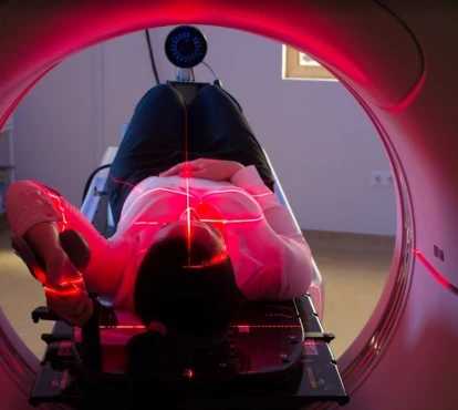
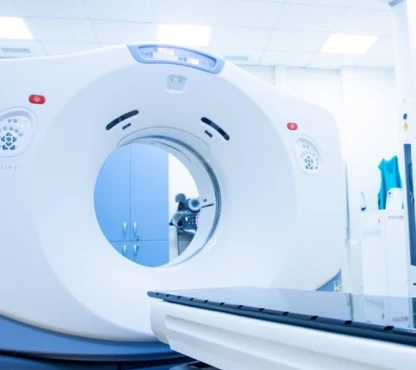
A comprehensive eye examination by an ophthalmologist, including a dilated fundus evaluation, is usually the initial step in diagnosing eye cancer. Various imaging tests, such as ultrasound, optical coherence tomography, and fluorescein angiography, are utilized to visualize and assess tumors within the eye. MRI and CT scans can also provide detailed images of the eye and surrounding structures. In some cases, a biopsy may be performed to obtain a tissue sample for histopathological analysis, which helps confirm the diagnosis and determine the tumor type.
Treatment Approaches [Weis et al., 2024]
What are the options for managing eye cancer?
Surgery:
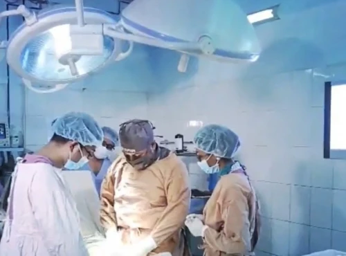
- Enucleation, the surgical removal of the entire eye, is often employed for large tumors or those that fail to respond to other treatments. This procedure may be necessary in cases of advanced ocular melanoma or retinoblastoma.
- Local tumor resection consists of tumor site-only removal, preserving the eye and vision, and is suitable for smaller tumors.
- Partial iris removal (iridectomy) may be an option for very small iris melanomas.
- Partial removal of the iris, along with a small piece of the outer eyeball (iridotrabeculectomy), is another potential treatment for small iris melanomas.
- Surgical removal of a portion of the iris and the ciliary body (iridocyclectomy) is used for small iris melanomas.
- Surgical removal of melanoma from the ciliary body or choroid (transscleral resection) is a complex procedure that should only be performed by experienced oncologists at specialized cancer centers, as it carries a high risk of damaging the rest of the eye and causing severe vision problems.
Radiation therapy options include:
- Plaque brachytherapy involves placing a radioactive disc near the tumor on the eye's surface to deliver targeted radiation. This treatment approach is frequently used for managing choroidal melanoma.
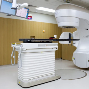
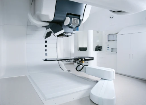
- External beam radiation therapy (EBRT) is a standard treatment for retinoblastoma and ocular lymphoma, where focused radiation beams are used to target the tumor. There are two primary versions of EBRT – proton beam radiation therapy (for large tumors and lesions besides the optic nerve) and stereotactic radiosurgery (Gamma Knife and CyberKnife tools).
The laser therapy method, called transpupillary thermotherapy, is a treatment that uses infrared light to heat and eliminate small choroidal melanoma tumors selectively.
Systemic chemotherapy, which uses drugs like vincristine, carboplatin, and etoposide, is a common treatment for retinoblastoma and ocular lymphoma. Additionally, direct injection of chemotherapeutic agents into the vitreous humor, known as intravitreal chemotherapy, is a treatment option for retinoblastoma.
Immunotherapy and targeted therapy:
- Pembrolizumab and nivolumab are immune checkpoint inhibitors used to treat metastatic ocular melanoma. These medications boost the immune system's ability to recognize and attack the cancer cells.
- Targeted therapies, such as selumetinib and tebentafusp, aim to treat ocular melanoma by targeting specific genetic mutations within the cancer cells. This personalized approach offers a tailored treatment option. Tebentafusp is a type of immunotherapy known as a bispecific T cell engager (BiTE). When it's injected into the body, one part of the drug binds to immune cells called T cells, while another part binds to a protein called gp100 on melanoma cells. This helps bring the T cells and cancer cells together, allowing the immune system to attack the cancer more effectively.
Prognosis & Follow-up
How does cutting-edge science improve the lifespan and quality of life for those with eye cancer?
The prognosis for eye cancer varies based on factors like the type, size, and location of the tumor, as well as whether the cancer has spread beyond the eye. For example, the 5-year survival rate for ocular melanoma that is confined to the eye is around 80%, but this rate declines significantly if the cancer has metastasized. Early diagnosis and prompt treatment are essential for achieving better outcomes.
Regular follow-up is crucial for assessing treatment effectiveness and identifying any cancer recurrence. Typical follow-up approaches include:
- Comprehensive eye examinations to check for signs of tumor reappearance or new growth.
- Periodic imaging tests, such as MRI or ultrasound, monitor for the spread of cancer to other areas of the body.
- Comprehensive health monitoring, including liver function tests, for patients with ocular melanoma, as the liver is a common site for metastatic spread.
Generally, first-year post-treatment follow-up is recommended every 3-4 months, from the second to fifth year – every six months, then – once a year.


