Introduction & Overview of Diffuse Large B-Cell Lymphoma
Diffuse Large B Cell Lymphoma (DLBCL) is a type of non-Hodgkin lymphoma (NHL), making up about one-third of all B cell NHL instances in the United States. Reports suggest there are 18,000 DLBCL cases annually [SEER].
This form of lymphoma can develop within or outside lymph nodes, impacting organs like the spleen, liver, and bone marrow. While DLBCL mainly affects individuals typically diagnosed around age 66, it can also manifest in children and young adults [Cancer Research UK].
DLBCL isn't an ailment but rather a cluster of cancers with two primary subcategories [Padala et al., 2023]:
-
- Germinal Center B-cell-like (GCB) DLBCL arises from B cells located in the centers of lymph nodes. Patients with this subtype often exhibit improved responses to chemotherapy treatments compared to ABC DLBCL – 83% vs 13%, respectively [Dunleavy and Wilson, 2014]. Prognostically, individuals with the GCB subtype generally fare better than those with the ABC subtype due to variances in the cancer cells' fundamental biology.
- Activated B-cell (ABC) DLBCL originates from B-cells transitioning into plasma cells, which is associated with a poor prognosis due to an overactive inflammatory molecule mediator called nuclear factor kappa B.
There are some common subtypes of DLBCL as per the ESMO Consensus;
-
- Primary Mediastinal B-cell Lymphoma (PMBCL) is typically seen in young adults and affects the chest area.
- CNS Lymphoma affects the brain and spinal cord.
- Transformed DLBCL can occur when a growing lymphoma transforms into a more aggressive form, such as follicular lymphoma (FL), chronic lymphocytic leukemia (CLL)/small lymphocytic lymphoma (SLL), or nodular lymphocyte-predominant Hodgkin lymphoma (NLPHL). These transformed lymphomas usually require more intensive types of treatment.
To confirm a diagnosis of DLBCL, doctors typically need to perform a biopsy by collecting a sample from the lymph node and examining it under a microscope. This procedure can be carried out under general anesthesia. Following the confirmation of diagnosis, determining the disease location in the body through staging becomes crucial since DLBCL is considered a type of blood cancer that may manifest across parts of the body [Cancer.net].
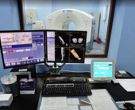
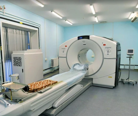
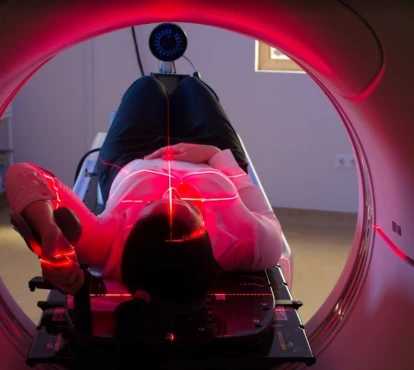
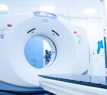
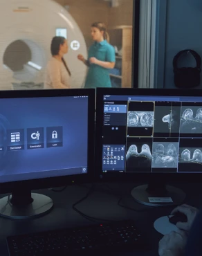
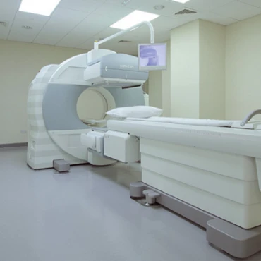
Doctors often conduct these tests using a positron emission tomography (PET) scan, which involves injecting a dye into the patient to pinpoint the location of cancer. Other diagnostic procedures may be part of the panel, such as a bone marrow biopsy to check for cancer in the bones or a spinal tap to examine the fluid around the brain and spinal cord for signs of cancer in the CNS system. The results from these tests help physicians determine the stage of lymphoma. The Ann Arbor staging system is utilized to categorize the extent of lymphoma, ranging from Stage I involving one lymph node or extralymphatic organ to Stage IV involving organs beyond lymph nodes.
Phases of Treatment
How is the treatment plan for DLBCL structured?
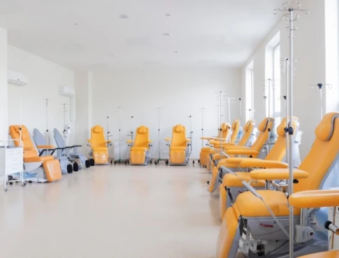
The treatment for DLBCL usually starts after the diagnosis. The goal is to achieve lasting remission or even a cure. A common approach involves using a mix of chemotherapy and a specific type of antibody that targets CD20, a protein in lymphoma cells. Rituximab (Rituxan) is an anti-CD20 antibody administered through intravenous infusion. For some patients, Rituxan Hycela, a form of rituximab injected under the skin, may also be considered [based on ESMO Guidelines].
Limited Stage (Stage) DLBCL
First Line Therapy
• The R-CHOP Regimen is the standard and the most common initial treatment, which consists of Rituximab, Cyclophosphamide, Doxorubicin, Vincristine, and Prednisone. This combination is typically given in cycles lasting 21 days each. Repeated for a total of 6 cycles.
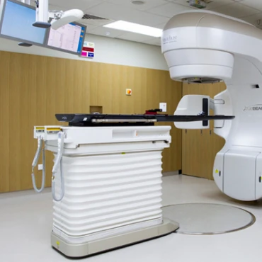
• Involved Site Radiotherapy (ISRT) - often combined with chemotherapy to improve control and outcomes in 60+ years patients with two relapse episodes or low-intermediate International Prognostic Index (IPI risk), which is a combination of stage III-IV of DLBCL, more than one extranodal sites involvement and / or slightly decreased performance status.
Intermediate Stage and Advanced Stage DLBCL
First Line Treatment
• R-CHOP or EPOCH-R chemotherapy. The standard approach for patients with the disease is R-CHOP, though high-risk patients might benefit from EPOCH R, which includes Etoposide and the standard R-CHOP medications in 6 cycles.
Second Line Treatment
• Salvage therapy for patients who relapse or do not respond to treatment includes Polatuzumab Vedotin combined with Bendamustine and Rituximab (BR) or dose chemotherapy followed by autologous stem cell transplant.
Recurrent or Treatment-resistant DLBCL
CAR T-Cell Therapy:
• Yescarta (Axicabtagene Ciloleucel) and Kymriah (Tisagenlecleucel) are the treatments that involve modifying the patient's T-cells to target the lymphoma cells and are used for patients who do not respond to at least two previous treatments. The CAR T-Cell Therapy may be combined with more intensive platinum-based chemotherapy regimens with cisplatin, gemcitabine, and high-dose cytarabine.
Nodular Lymphocyte Predominant Hodgkin Lymphoma (NLPHL)
• Observation Approach: Sometimes treatment may not be needed for NLPHL, so doctors opt for a waiting strategy. This involves check-ups and starting treatment only if the condition worsens.
Targeted Treatment
• Adcetris (Brentuximab Vedotin) is used in cases where NLPHL recurs, combining therapy with chemotherapy.
• Opdivo (Nivolumab) and Keytruda (Pembrolizumab) help boost the system's response to cancer cells, and they are being studied in terms of types of lymphomas.
Assessing Response
Evaluating how well the treatment works involves repeated PET scans to see if the lymphoma has shrunk or become less active. The aim is remission (CR), which means there are no signs of the disease.
Prognosis, Survival Rates, and Monitoring
What are the chances of survival, and what factors influence outlook?
The five-year survival rate varies depending on the stage of the disease and other factors. Generally, it ranges from 60-70% for early-stage disease to 50% for advanced stages.
Moreover, data on outcomes provided by the National Cancer Institute indicate a correlation with age, showing a prognosis of 80% for patients under 55, 70% for those aged 55-64, and 55% for individuals over 65 [SEER].
Consistent monitoring is essential to detect relapses and address long-term side effects, as highlighted by the Lymphoma Research Foundation:
1. Initial check-ups: Typically scheduled every three months during the two years after treatment involving examinations and necessary imaging.
2. Ongoing follow-up visits every six months from years three to five, and then after that to address survivorship concerns and screen for potential secondary cancers.

