Introduction
Ewing’s sarcoma is a type of bone tissue tumor affecting mostly children and young adults. This tumor gets its name from Dr. James Ewing, the doctor who first described it in the 1920s. Later experts found this same type of tumor in soft tissues and called it extraosseous (outside of the bone) Ewing tumor. Another rare childhood cancer that's a lot like these sarcomas is called peripheral primitive neuroectodermal tumor (PNET). It can also start in the bone or soft tissues. These tumors are now all referred to as the Ewing family of tumors or Ewing sarcomas. They are all treated in similar ways.
Ewing sarcoma rarely occurs in children under 5 years, the peak incidence is between 10 and 15 years. This is associated with puberty - the period of active growth of skeletal bones. The most common places for Ewing sarcoma to start include the:
- Bones in the chest wall, like the ribs or shoulder blades
- Hip bones (pelvis)
- Leg bones (mostly in the middle of the long bones)
Extraosseous Ewing tumors can start almost anywhere in the body.
The tumor usually grows into the tissue surrounding the bone, destroying it in the process. The cancer cells can also spread to other parts of the body very quickly. This process is called metastasis. Approximately 25% of patients have metastasis at the time of diagnosis. In about half of them, the disease spreads to the lungs.
Etiology
No one knows exactly why children get Ewing's sarcoma. Neither any external factors influencing the body (for example, when the child previously received radiation therapy), nor any hereditary genetic factors (hereditary predisposition) are of great importance.
It is known that Ewing's sarcoma cancer cells have certain chromosomal changes. These changes always affect a specific gene on chromosome 22. This gene is called the Ewing sarcoma gene (you can also find its other name EWS gene). As a result, when changes lead to gene damage, it causes a healthy cell to become cancerous. But in principle, all such gene changes that are found in tumor tissue are not inherited.
Signs and Symptoms
Depending on the location of the tumor a variety of symptoms may be reported. The most common for all tumors are:
- pain and swelling because of tumor growth and tissue destruction;
- tenderness to palpation of the affected area;
- impairment of function of the affected area: gait disorder, claudication, decreased range of motion of the limb;
- bone fractures without obvious mechanical impact;
- tumor intoxication: fever, rapid fatigability, body weight loss.
Symptoms depend on the location of the tumor. For example, when the vertebrae are affected, the first symptom may be compression of the spinal cord (paralysis, pelvic floor dysfunction).
Pain is usually the first symptom and is often associated with physical activity, later pain becomes constant, disturbs night sleep and requires painkillers.
Diagnosis
The examination usually begins with visualization of the affected area of the body: an X-ray may be done to see if a tumor is present.
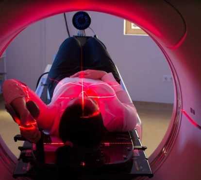
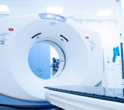
Other tests administered to obtain an image of the tumor, its size and spread beyond the bone include:
- a CT (computerized tomography) scan;
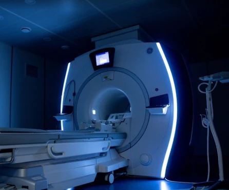
- an MRI (magnetic resonance imaging) scan;
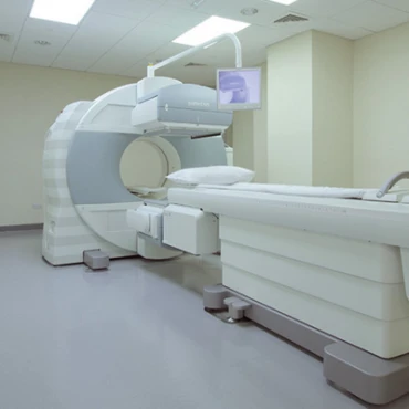
- a bone scan (scintigraphy) or total body PET-CT (positron emission tomography and computed tomography).
These imaging tests are enhanced with contrast which helps to detect areas of increased or decreased bone metabolism and can identify abnormal processes involving the bone, including a tumor.
To estimate the tumor spread the doctor may perform the bone marrow harvest.
To definitively confirm the diagnosis of Ewing's sarcoma, it is necessary to take a sample of tumor tissue (biopsy). The biopsy should only be performed by specialists who will subsequently perform sarcoma surgery, or who have experience in sarcoma surgery. Doctors use various cell studies to verify the tumor process: histologic study, immunohistochemistry assay, cytogenetic analysis for the presence of the t(11;22) translocation and other chromosomal changes, molecular diagnostic (study of the genetic code of the tumor).
Routine laboratory tests are also carried out. Changes in the clinical blood test may be observed: leukocytosis, anemia, increased ESR. In a biochemical blood test, the level of lactate dehydrogenase (LDH) is important, since its increase is more often observed with a more aggressive course of the disease.
A general examination is necessary to determine the body's ability to tolerate treatment.
Treatment
Treatment of Ewing's sarcoma includes a combination of several treatment methods: chemotherapy, surgery and radiation therapy. The main goal of treatment is to destroy not only visible tumor foci, but also those tumor cells that cannot yet be seen during examination. This is why treatment of Ewing's sarcoma should be systemic, not just local.
Neoadjuvant Chemotherapy
Сhemotherapy, which is carried out before the local treatment (surgery or radiotherapy) is called neoadjuvant or induction. Neoadjuvant chemotherapy can fulfill three key functions:
● it can completely eradicate cancer cells, offering a potential cure;
● it can control the growth and spread of the disease;
● it can alleviate symptoms by reducing the size of a tumor and make the next local step less traumatic.
Commonly induction chemo consists of several repeating courses that include several cytostatics (drugs that destroy tumor cells). [2] They can be a combination of vincristine, doxorubicin, cyclophosphamide and ifosfamide, etoposide.[3] Cytostatic drugs can cause a decrease in healthy blood cells and your doctor can prescribe you special medicine for bone marrow stimulation. It is called filgrastim.
The chemotherapy period is dangerous for the development of infectious complications and bleeding. To avoid complications, you need to follow your doctor's instructions.
Local therapy
Local treatment is usually incorporated after initial 5-6 cycles of chemotherapy and if there is good response to chemotherapy in both local site and metastatic site.
The choice of local treatment is influenced by multiple factors, such as age of the patients, site and size of the tumor, local extent of the tumor, clinic-radiological response to chemotherapy, expertise and experience of the treating institution and surgeon and patient’s choice. The main types of local treatment are surgery and radiotherapy.
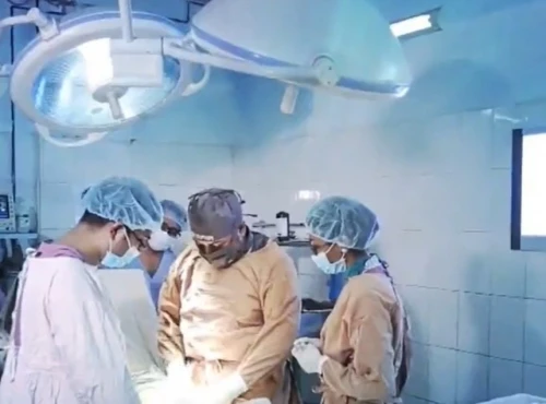
The surgical resection principle depends on the respectability, size and sites of the tumor and its operability after chemotherapy. Surgical resection should be tried whenever a marginal or wide resection is feasible. When complete removal of the tumor is impossible, you should think about radiation therapy or a combination of surgery and radiation therapy. Currently, most surgical interventions preserve the affected limb and its functionality, including through prosthetics. But sometimes an amputation is necessary.
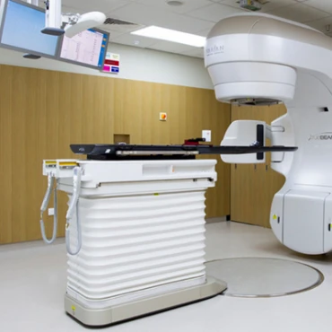
The main principle of radiotherapy is to avoid toxicity to adjacent normal organs which is especially important when conducting radiation therapy in the pelvic and chest areas.
Local Therapy for Metastatic Sites
The most common metastasis of Ewing's sarcoma is the lungs. Bilateral pulmonary irradiation at a dose of 14–20 Gy has been reported to improve the outcome of patients with pulmonary disease. It is usually carried out at the end of the consolidation stage or in parallel with it.
Adjuvant (Postoperative) Chemotherapy
Further therapy depends on the spread of the disease and the effectiveness of the treatment. In removed tumor we can evaluate the pathomorphosis (number of viable tumor cells). IV-degree therapeutic pathomorphosis is also called pathological complete response. This means that most of the tumor cells have died due to induction chemotherapy and we can repeat eight cycles of the same drugs in the adjuvant chemotherapy.
In other cases when the therapeutic pathomorphosis is III degree or less we should change treatment plan and choose a regimen with high-dose chemotherapy followed by autologous stem cell transplantation or novel agents therapy. When the degree of pathomorphosis cannot be studied, response to treatment is assessed by a decrease in the volume of the primary tumor and the absence of new metastatic foci:
● complete response - complete regression of the soft tissue component of the tumor, absence or disappearance of distant metastases, disappearance of periosteal reaction, reduction of osteolytic foci according to X-ray, CT and MRI studies;
● partial response - reduction of more than 50% of the tumor mass compared to initially diagnosed volume;
● stable disease - reduction less than 50% or progression less than 25% of initial volume of tumor lesion;
● disease progression - progression more than 25% of the initial volume lesions or the appearance of new metastases.
For patients with stable disease and partial response it is necessary to escalate treatment: administer high-dose chemotherapy or admit novel agents or increase the number of courses of adjuvant chemotherapy.
Patients with disease progression have poor prognosis, we can try to change cytostatics but in case of further progression, patients require palliative therapy.
Novel Treatment Options
At the moment, no new treatment methods with a clear positive effect have been found. A complete analysis of the tumor genome can identify points of application for targeted therapy. We should perform this kind of examination in all patients with metastatic disease or tumor progression.
Prognosis
The survival rates for Ewing sarcoma vary based on several factors. These include the stage of tumor, a person’s age and general health, and how well the treatment plan works. There are other factors that have been linked to higher survival rates for all stages of Ewing tumors. These factors include being younger than 10, having a smaller sized tumor, having a tumor located in an arm or leg, and having normal levels of the enzyme lactate dehydrogenase (LDH).
The overall 5-year relative survival rate for people with Ewing sarcoma is 63%. If the tumor is found only in the area it began (called localized), the 5-year relative survival rate is 82%. If it has spread to the nearby region (called regional), the 5-year relative survival rate is 71%. If the tumor has spread to distant areas at the time of diagnosis (called metastasis), the 5-year relative survival rate is 39%.


