Epidemiology & Classification of Acute Lymphocytic Leukemia
Acute Lymphocytic Leukemia (ALL) is a type of blood cancer originating in the bone marrow and primarily affecting white blood cells known as lymphocytes. ALL can occur at any age, but it is most commonly – around 80% - diagnosed in children, with a peak incidence between ages 2 and 5. ALL is less common in adults but tends to have a more challenging prognosis. The median age of diagnosis for adults is around 35-50 years old [cancer.gov].
ALL is classified into several subtypes based on the type of lymphocytes affected and specific genetic markers [American Society of Hematology, 2024]. Here are the main subtypes:
- B-cell ALL: This is the most common subtype, accounting for 75-80% of ALL cases in children and adults. B-cell ALL originates from immature B-lymphocytes. B-cell ALL often involves specific genetic mutations such as the t(12;21) translocation, associated with a better prognosis.
- T-cell ALL: This subtype accounts for 15-20% of ALL cases. It originates from immature T-lymphocytes and is more common in older children and adolescents. T-cell ALL presents with a high white blood T-cell count and often involves the thymus, leading to a mediastinal mass.
- Philadelphia Chromosome-positive (Ph+) ALL: This subtype is characterized by the presence of the Philadelphia chromosome, a result of the t(9;22) translocation. Ph+ ALL is more common in adults and requires targeted therapy with tyrosine kinase inhibitors (TKIs) in addition to standard chemotherapy.
- Mixed Lineage Leukemia (MLL) rearranged ALL: This subtype is associated with rearrangements in the MLL gene, which can occur in both B-cell and T-cell ALL. MLL-rearranged ALL is more common in infants and is associated with a poorer prognosis.
ALL is typically classified based on the presence and spread of leukemia cells. Although ALL does not have a formal staging system like solid tumors, it is categorized by phases of treatment and the disease's extent at diagnosis:
- Stage I: This stage involves the presence of leukemia cells in the bone marrow and blood without spreading to other parts of the body. The goal at this stage is to achieve remission through induction therapy.
- Stage II: In this stage, leukemia cells may be found in the central nervous system (CNS) or other extramedullary sites. CNS prophylaxis or treatment is essential to prevent or manage the spread of leukemia cells to the brain and spinal cord.
- Stage III: At this stage, leukemia cells have spread to other organs and tissues beyond the bone marrow and blood. This may include the liver, spleen, and lymph nodes. Intensive consolidation therapy is used to eliminate remaining leukemia cells.
- Stage IV: This stage is characterized by a high burden of leukemia cells in multiple organs and tissues, including the bone marrow, blood, CNS, and other extramedullary sites. Treatment involves aggressive therapy, including potential stem cell transplantation and targeted therapies to achieve and maintain remission.
Algorithm of Diagnosis
What evaluations do ALL patients undergo to identify the best treatment strategy?
Diagnosing ALL involves a series of evaluations to determine the disease's stage and clinical presentation, including [National Comprehensive Cancer Network, 2021]:
- Clinical Examination includes a thorough physical exam where the doctor checks for signs of anemia, infections, and bleeding. The examination often involves palpating the abdomen to check for an enlarged liver or spleen and inspecting lymph nodes.
- Blood Tests: A complete blood count (CBC) measures the levels of red cells, white cells, and platelets. In ALL, there are typically high numbers of immature white blood cells (blasts) and low levels of red cells and platelets.
- Bone Marrow Examination: A definitive diagnosis of ALL requires a bone marrow aspiration and biopsy. These procedures involve extracting bone marrow fluid and tissue, usually from the hip bone, to examine under a microscope for the presence of leukemia cells.
- Cytogenetic and Molecular Tests: These tests analyze the chromosomes of the leukemia cells to identify specific genetic abnormalities. For B-cell ALL, common mutations include t(12;21) and hyperdiploidy. In T-cell ALL, common genetic changes include mutations in the NOTCH1 gene. Ph+ ALL is defined by the BCR-ABL1 fusion gene resulting from the t(9;22) translocation. MLL-rearranged ALL involves the MLL gene rearrangements.
- Lumbar Puncture: Also known as a spinal tap, this test checks for the presence of leukemia cells in the cerebrospinal fluid, indicating central nervous system involvement.
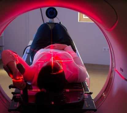
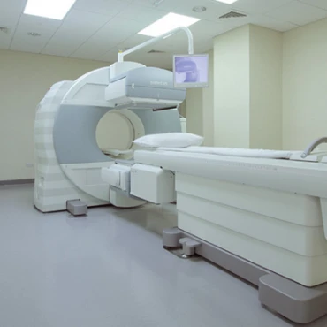
- Imaging Studies: CT scans, MRIs, or ultrasounds may be used to detect any enlarged organs or lymph nodes and evaluate the disease's spread.
Phases of Treatment
How is ALL treatment structured?
Treatment for ALL is divided into several phases, each designed to target leukemia cells at different stages and prevent relapse [cancer.org]. The main phases are:
1. Induction Therapy
The goal of induction therapy is to achieve remission by reducing the number of leukemia cells to undetectable levels. This phase typically lasts about four weeks and involves intensive chemotherapy, such as vincristine, daunorubicin, prednisone, and asparaginase. Around 80% of adults and 95% of children with ALL achieve remission after one month of induction therapy.
2. Consolidation (Intensification) Therapy
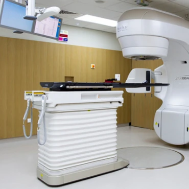
Following remission, consolidation therapy aims to eliminate any remaining leukemia cells. This phase often involves higher doses of chemotherapy drugs and may include additional treatments such as radiation therapy or targeted therapies (imatinib, dasatinib). The duration of consolidation therapy can vary but usually lasts several months.
3. Maintenance Therapy
Maintenance therapy helps keep the leukemia in remission and typically lasts for 2-3 years. It involves lower doses of chemotherapy drugs, often taken orally, and periodic intravenous treatments. Common maintenance drugs include methotrexate and 6-mercaptopurine.
First-Line Treatment Options
What are the initial treatments for ALL?
Chemotherapy is the cornerstone of ALL treatment. The specific drugs and regimens used depend on the patient's age, overall health, and specific genetic markers of the leukemia cells. Induction chemotherapy often includes a combination of drugs such as vincristine, daunorubicin, cyclophosphamide, and prednisone. Based on the patient's prognostic factors – younger than 50 years old, with white blood cell count < 30,000 for B-cell ALL and <100,000 for T-cell ALL - some regimens may also include cyclophosphamide, L-asparaginase (or pegaspargase), and/or high doses of methotrexate or cytarabine (ara-C) as part of the induction phase [cancer.org].
Targeted Therapy: For patients with Philadelphia chromosome-positive (Ph+) ALL, targeted therapy with tyrosine kinase inhibitors (TKIs) such as imatinib or dasatinib is combined with chemotherapy. These drugs target the abnormal protein produced by the Philadelphia chromosome, which drives the growth of leukemia cells [ashpublications.org].
Central Nervous System (CNS) Prophylaxis: Leukemia cells can hide in the central nervous system, so CNS prophylaxis is a critical component of ALL treatment. This often involves intrathecal chemotherapy, where drugs (cytarabine, methotrexate) are injected directly into the cerebrospinal fluid, and sometimes cranial radiation therapy.
Second-Line Treatment Options
What options are available if initial treatments fail?
Stem Cell Transplantation: For patients with high-risk ALL or those who relapse, an allogeneic stem cell transplant (SCT) is a crucial treatment option. This involves high-dose chemotherapy to eradicate leukemia cells, followed by a donor's infusion of healthy stem cells. SCT can significantly improve survival rates but has substantial risks and side effects [jnccn.org].
Monoclonal Antibodies: Newer treatments include monoclonal antibodies like blinatumomab and inotuzumab ozogamicin. Blinatumomab is a bispecific T-cell engager (BiTE) that directs the patient's T-cells to attack leukemia cells, while inotuzumab ozogamicin delivers a cytotoxic agent directly to leukemia cells.
CAR-T Cell Therapy: Chimeric Antigen Receptor (CAR) T-cell therapy is an advanced treatment where a patient's T-cells are genetically modified to attack leukemia cells. This therapy has shown promising results in patients with refractory or relapsed ALL but is only available in specialized centers.
Prognosis and Survival Rates
What are the survival rates and factors affecting prognosis?
The prognosis for ALL varies significantly based on several factors, including the patient's age, overall health, genetic characteristics of the leukemia cells, and the patient's response to initial treatment [seer.cancer.gov]. Patients who go into complete remission (no visible leukemia in the bone marrow – see below) within 4 to 5 weeks of starting treatment tend to have a better prognosis than those for whom this takes longer. Key statistics on prognosis predictions include:
- Overall Survival Rate: The 5-year survival rate for adults with ALL is approximately 35-40%, but this can be higher in younger adults and those who achieve complete remission after induction therapy.
- Children: The prognosis for children with ALL is much better, with 5-year survival rates exceeding 90% due to more aggressive and effective treatment protocols.
- High-Risk Factors: Factors such as the presence of the Philadelphia chromosome, high white blood cell count at diagnosis, and older age can negatively impact the prognosis.


