Definition
Aortic dissection or aortic dissecting aneurysm is a defect in the inner membrane of aneurysmal dilated aorta, accompanied by hematoma formation, longitudinally dissectingthevascular wall with the formation of a false channel. Aortic dissecting aneurysm is manifested by sudden intense pain migrating along the course of the dissection, elevated blood pressure, signs of ischemia of the heart, brain and spinal cord, kidneys, and internal bleeding. The diagnosis of vascular wall dissection is based on echocardiography, CT, MRI of the thoracic/abdominal aorta, and aortography. Treatment of complicated aneurysm includes intensive drug therapy and resection of the damaged part of the aorta with subsequent reconstructive repair.
General information
Aortic dissecting aneurysm is a longitudinal dissection of the aortic wall distal or proximal to the aorta at various lengths caused by the rupture of its inner membrane and blood penetration into the thickness of the degeneratively altered middle layer. Aortic dilatation during aortic wall dissection may be moderate or absent. Therefore, aortic dissection aneurysm is often referred to as aortic dissection.
Most aneurysms are localized in the most hemodynamically vulnerable areas of the aorta: about 70% – in the ascending aorta a few centimeters from the aortic valve, 10% of cases – in the arch, 20% – in the descending aorta distal to the orificeof the left subclavian artery. Dissecting aneurysms in cardiology refers to life-threatening conditions with the risk of massive bleeding in case of aortic rupture or acute ischemia of vital organs (heart, brain, kidneys, etc.) in case of occlusion of the main arteries. Aortic aneurysm dissection usually occurs at the age of 60-70 years in men, 2-3 times more often than in women.
Causes
The causes of pathology are diseases and conditions leading to degenerative changes in the muscular and elastic structures of the middle aortic sheath (mediastinum). Older age of patients (older than 60-70 years), chest trauma, and third trimester of pregnancy in women older than 40 years are considered risk factors for aortic aneurysm dissection. Underlying causes include:
- Elevated BP. The main risk of aortic dissection is associated with prolonged arterial hypertension (70-90% of cases), accompanied by hemodynamic stress and chronic aortic traumatization.
- Hereditary defects of connective tissue. A dissecting aneurysm may develop as a complication of Marfan, Turner, and Ehlers-Danlos syndromes.
- Heart and vascular diseases. The risk group includes patients with aortic malformations, coarctation of the aorta, severe atherosclerosis of the aorta, and systemic vasculitis.
- Postponed cardiac surgical operations and manipulations. In the early and late postoperative period after cardiac and aortic surgery (aortic valve replacement, aortic resection), there is an increased risk of aneurysm dissection. Iatrogenic dissecting aneurysms are associated with technical errors during aortography, balloon dilatation, and cannulation of the aorta to provide artificial circulation.
Pathogenesis
The primary pathogenetic link in most cases is an intimal rupture with subsequent formation of an intramural hematoma. In about 10% of cases, aortic dissecting aneurysm may be initiated by mediastinal hemorrhage from spontaneous rupture of capillaries branching in the aortic wall. Dissemination of intramural hematoma within the media is usually accompanied by subsequent intimal rupture but may occur without it (in 3-13% of cases). In rare cases, aortic dissection may be observed at penetration of atherosclerotic ulcer.
Classification
According to DeBakey’s classification, three types of stratification are defined:
- I – intimal tear in the ascending segment of the aorta; dissection extends to the thoracic and abdominal sections;
- II – the site of rupture and dissection is limited to the ascending aorta,
- III – intimal tear in the descending aorta, dissection may extend to the distal abdominal aorta, sometimes retrogradely to the arch and ascending part.
The Stanford classification distinguishes type A aortic dissecting aneurysms, with proximal dissection involving the ascending aorta, and type B, with distal dissection of the aortic arch and descending aorta. Type A is characterized by a higher incidence of early complications and high pre-hospital mortality. Aortic dissecting aneurysms can be acute (from several hours to 1-2 days), subacute (from several days to 3-4 weeks), and chronic (several months).
Symptoms
The clinical picture of the disease is determined by the presence and extent of aortic dissection, intra-wall hematoma, compression and occlusion of aortic branches, and ischemia of vital organs. There are several variants of aortic dissecting aneurysm development: formation of extensive unruptured hematoma; wall dissection and hematoma breakthrough into the aortic lumen; wall dissection and hematoma breakthrough into the surrounding aortic tissues; aortic rupture without wall dissection.
Aortic dissection is characterized by a sudden onset with imitation of symptoms of various cardiovascular, neurological, and urological diseases. Aortic dissection is manifested by a sharp increase in tearing, intolerable pain with a wide area of irradiation (behind the sternum, between the shoulder blades and along the spine, in the epigastric region, lumbar region), migrating along the course of the dissection. There is an increase in blood pressure with subsequent decline, asymmetry of pulse on the upper and lower extremities, profuse sweating, weakness, lividity, and motor restlessness. The majority of patients with aortic dissecting aneurysms die from the development of complications.
Neurological manifestations of pathology may include ischemic lesions of the brain or spinal cord (hemiparesis, paraplegia), peripheral neuropathy, and disorders of consciousness (syncope, coma). A dissecting aneurysm of the ascending aorta may be accompanied by myocardial ischemia, compression of mediastinal organs (hoarseness, dysphagia, dyspnea, Horner syndrome, superior vena cava syndrome), development of acute aortic regurgitation, hemopericardium, and cardiac tamponade.
Diagnosis
If a patient is suspected of having an aortic dissection aneurysm, an urgent and accurate assessment of the patient’s condition is necessary. The main diagnostic methods for visualizing aortic lesions are chest radiography, echocardiography (transthoracic and transesophageal), ultrasound, MRI, and CT of the thoracic/abdominal aorta, and aortography.
- Chest radiography reveals signs of spontaneous aortic dissection: dilation of the aorta and upper mediastinum (in 90% of cases), deformation of the shadow of the aortic or mediastinal contours, presence of pleural effusion (more often on the left), and decreased or absent pulsation of the dilated aorta.
- Echocardiography. Transthoracic or transesophageal echocardiography helps determine the state of the thoracic aorta, identify detached intima flaps and true and false channels, assess the aortic valve, and determine the prevalence of atherosclerotic lesions of the aorta.
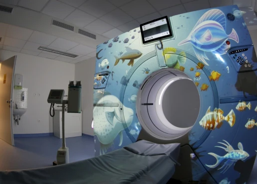
- Tomography. CT and MRI for dissecting aortic aneurysm require the stable condition of the patient for transportation and the procedure. CT is used to detect intramural hematoma and penetration of atherosclerotic ulcers of the thoracic aorta. MRI allows, without the use of intravenous injection of contrast agent, to accurately determine the localization of intimal rupture, the direction of dissection in the direction of blood flow in the false channel, to assess the involvement of the main branches of the aorta, the state of the aortic valve.
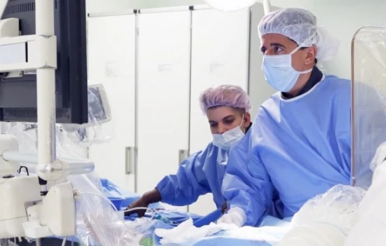
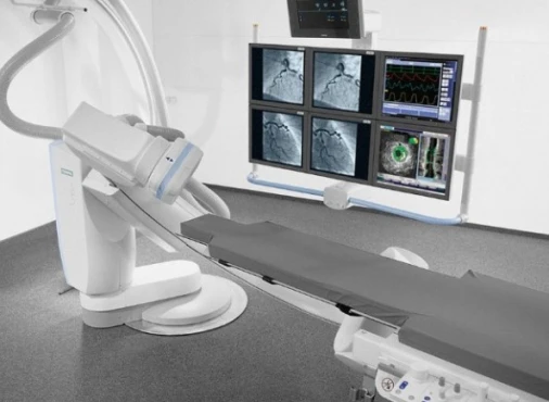
- Aortography is an invasive but highly sensitive method of investigating aortic dissecting aneurysms; it allows the site of initial rupture, localization and extent of dissection, true and false lumen, presence of proximal and distal fenestration, degree of aortic valve and coronary artery integrity, and integrity of aortic branches to be seen.
It is necessary to perform differential diagnosis of aortic dissecting aneurysm with acute myocardial infarction, mesenteric vessel occlusion, renal colic, renal infarction, thromboembolism of the aortic bifurcation, acute aortic insufficiency without aortic dissection, non-dissecting aneurysm of the thoracic or abdominal aorta, stroke, mediastinal tumor.
Treatment of aortic dissection
Patients with complicated aortic aneurysms are urgently hospitalized in the cardiac surgery department. Conservative therapy is indicated in any form of the disease at the initial stage of treatment to stop the progression of vascular wall dissection and stabilize the patient’s condition. Following treatment options are conducted most often:
- Intensive therapy aims to relieve pain (by administering non-narcotic and narcotic analgesics), remove the patient from shock, and reduce blood pressure. Hemodynamics, heart rate, diuresis, CVD, and pulmonary artery pressure are monitored. In clinically significant hypotension, it is essential to restore blood volume quickly by intravenous infusion of solutions.
- Medical treatment. It is the primary treatment in most patients with uncomplicated dissecting aneurysms of type B (distal dissection), stable isolated aortic arch dissection, and stable uncomplicated chronic dissection. In case of ineffectiveness of the current therapy, progression of dissection, and development of complications, as well as in patients with acute proximal aortic wall dissection (type A), emergency surgical intervention is indicated immediately after stabilization of the condition.
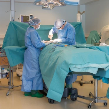
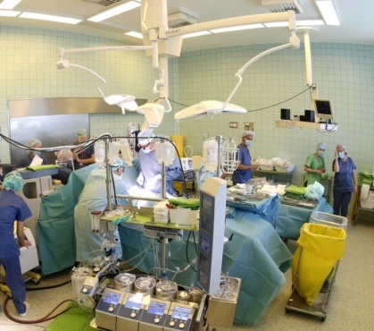
- Operative treatment. In an aortic dissecting aneurysm, the damaged part of the aorta with rupture is resected, the intimal flap is removed, the false lumen is eliminated, and the dissected aortic fragment is reconstructed (sometimes simultaneously, several aortic branches are reconstructed) by prosthesis or convergence of the ends. In most cases, the operation is performed under artificial circulation. If indicated, valvuloplasty or aortic valve prosthesis and reimplantation of coronary arteries are performed.
Prognosis and prevention
Untreated aortic dissecting aneurysms have a high mortality rate, which can be as high as 90% during the first three months. The postoperative survival rate for type A dissection is 80%, and for type B, it is 90%. Long-term prognosis is generally favorable: ten-year survival rate is 60%. Prevention of aortic dissecting aneurysm formation consists of controlling the course of cardiovascular disease. Prevention of aortic dissection includes observation by a cardiologist, blood pressure and cholesterol levels monitoring, and periodic aorta ultrasound.


