Introduction
A thoracoabdominal aortic aneurysm is a pathologic local expansion of a section of the main artery throughout its entire length due to the weakness of its walls. Depending on the localization, an aortic aneurysm may manifest with pain in the chest or abdomen, the presence of a pulsating tumor-like mass, and symptoms of compression of adjacent organs: dyspnea, cough, dysphonia, dysphagia, swelling, and cyanosis of the face and neck. The basis of aortic aneurysm diagnosis consists of radiological (chest and abdominal radiography, aortography) and ultrasound methods (duplex scanning of the thoracic/abdominal aorta). Surgical treatment of the aneurysm involves its resection with aortic prosthesis or closed endoluminal prosthesis of the aneurysm with a special endoprosthesis.
General information
Aortic aneurysm is characterized by irreversible dilation of the lumen of the arterial trunk in a limited area. The ratio of aortic aneurysms of different localization is approximately as follows: abdominal aortic aneurysms account for 37% of cases, ascending aorta - 23%, aortic arch - 19%, descending thoracic aorta - 19.5%. Thus, thoracic aortic aneurysms in cardiology account for almost 2/3 of all pathology. Thoracic aortic aneurysms are often combined with other aortic malformations - aortic insufficiency and coarctation of the aorta.
Causes
By etiology, all aortic aneurysms can be divided into congenital and acquired. The formation of congenital aneurysms is associated with hereditary diseases of the aortic wall:
- Marfan syndrome
- fibrous dysplasia
- Ehlers-Danlos syndrome.
- Erdheim's syndrome
- hereditary elastin deficiency, etc.
Acquired aortic aneurysms can have inflammatory and non-inflammatory etiologies:
- Postinflammatory aneurysms result from specific and nonspecific aortitis in fungal lesions of the aorta, syphilis, and postoperative infections.
- Non-inflammatory degenerative aneurysms are due to atherosclerosis, defects in suture material, and aortic prostheses.
- Hemodynamic-post stenotic and traumatic aneurysms are associated with mechanical injury to the aorta.
- Idiopathic aneurysms develop in aortic medionecrosis.
Older age, male gender, arterial hypertension, tobacco smoking and alcohol abuse, and hereditary aggravation are considered risk factors for the formation of aortic aneurysms.
Pathogenesis
In addition to aortic wall defects, mechanical and hemodynamic factors are involved in aneurysm formation. Aneurysms are more likely to occur in functionally stressed areas under increased stress due to high blood flow velocity, steepness of the pulse wave, and its shape. Chronic traumatization of the aorta and increased activity of proteolytic enzymes destroy the elastic framework and cause nonspecific degenerative changes in the vessel wall.
A formed aortic aneurysm progressively increases in size as the stress on its walls increases in proportion to the expansion of its diameter. Blood flow in the aneurysmal sac slows down and becomes turbulent. Only about 45% of the blood volume in the aneurysm enters the distal arterial bed. This is because blood rushes along the walls when entering the aneurysmal cavity, and the turbulence mechanism and the presence of thrombotic masses in the aneurysm restrain the central flow. Thrombus in the aneurysm cavity is a risk factor for thromboembolism of distal aortic branches.
Classification
Several classifications of aortic aneurysms have been proposed in vascular surgery, considering their localization by segments, shape, wall structure, and etiology. In accordance with the segmental classification, the following are distinguished
- aneurysm of Valsalva sinuses
- ascending aortic aneurysm
- aortic arch aneurysm
- descending aortic aneurysm
- abdominal aortic aneurysm
- aneurysm of combined localization - thoracoabdominal aneurysm.
Evaluation of the morphologic structure of aortic aneurysms allows us to subdivide them into true and false (pseudoaneurysms):
- A true aneurysm is characterized by thinning and bulging outward of all aorta layers. The etiology of true aortic aneurysms is usually atherosclerotic or syphilitic.
- Pseudoaneurysm. The wall of a false aneurysm is represented by connective tissue formed due to the organization of a pulsating hematoma; the native aortic walls are not involved in forming a false aneurysm. By origin, they are more often traumatic and postoperative.
Considering the clinical course, uncomplicated, complicated, and dissecting aortic aneurysms are distinguished. Specific complications of aortic aneurysms include ruptures of the aneurysmal sac accompanied by massive internal bleeding and hematoma formation, aneurysm thrombosis and arterial thromboembolism, and phlegmons of the surrounding tissues due to infection of the aneurysm.
A particular type is a dissecting aortic aneurysm, when, through a rupture of the inner membrane, blood penetrates between the layers of the arterial wall and spreads under pressure along the course of the vessel, gradually dissecting it.
Symptoms of an aortic aneurysm
Clinical manifestations of aortic aneurysms are variable and determined by the localization, size of the aneurysmal sac, extent, and etiology of the disease. Aneurysms may be asymptomatic or accompanied by scanty symptomatology and detected during preventive examinations. The leading manifestation is pain due to the lesion of the aortic wall, its stretching or compression syndrome.
Abdominal aortic aneurysm
The clinic of abdominal aortic aneurysm is manifested by transient or persistent diffuse pain, abdominal discomfort, belching, heaviness in the epigastrium, sensation of gastric overflow, nausea, vomiting, bowel dysfunction, and weight loss. Symptomatology may be associated with compression of the cardiac section of the stomach, the duodenum, and the involvement of visceral arteries. Often, patients independently determine the presence of increased pulsation in the abdomen. On palpation, a tense, dense, painful pulsating mass is detected.
Thoracic aortic aneurysm
Aortic aneurysm of the ascending aorta is characterized by pain in the heart region or behind the sternum due to compression or stenosis of the coronary arteries. Patients with aortic insufficiency are bothered by dyspnea, tachycardia, and dizziness. Large aneurysms cause the development of superior vena cava syndrome, which includes headaches and swelling of the face and upper half of the trunk.
Aortic arch aneurysm leads to esophageal compression with dysphagia; in case of compression of the recurrent nerve, there is voice hoarseness (dysphonia), dry cough; vagus nerve involvement is accompanied by bradycardia and salivation. In compression of the trachea and bronchi, dyspnea and stridor breathing develop; in compression of the lung root - congestion and frequent pneumonia.
Pain in the left arm and scapula occurs when a descending aortic aneurysm irritates the periaortic sympathetic plexus. If the intercostal arteries are involved, spinal cord ischemia, paraparesis, and paraplegia may develop.
Thoracoabdominal aortic aneurysm
This type of aortic aneurysm is characterized by a combination of abdominal and thoracic symptoms, so the clinical presentation is usually more varied. In addition, this type of aneurysm increases the likelihood of life-threatening complications such as rupture or dissection of the aneurysm.
Complications
A rupture with massive bleeding, collapse, shock, and acute heart failure may complicate aortic aneurysms. Aneurysm rupture can occur in the superior vena cava system, pericardial and pleural cavity, esophagus, and abdominal cavity. This leads to severe, sometimes fatal conditions - superior vena cava syndrome, hemopericardium, cardiac tamponade, hemothorax, and pulmonary, gastrointestinal, or intra-abdominal bleeding.
When thrombotic masses detach from the aneurysmal cavity, a picture of acute occlusion of the limb vessels develops cyanosis, pain in the toes, and intermittent claudication. Renovascular arterial hypertension and renal failure occur in renal artery thrombosis; stroke occurs in cerebral artery lesions.
Diagnosis
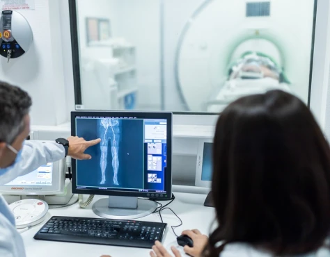
Diagnostic search for aortic aneurysm includes assessment of subjective and objective data, radiologic, ultrasound, and tomographic studies. An auscultatory sign of aneurysm is the presence of a systolic murmur in the projection of aortic dilation. Abdominal aortic aneurysms are detected by palpation of the abdomen in the form of a tumor-like pulsating formation. Following diagnostic tests are usually used:
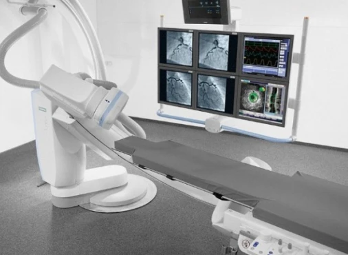
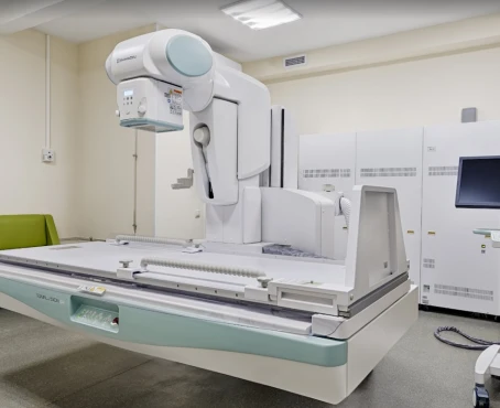
- Radiography. The plan of radiological examination of patients with thoracic or abdominal aortic aneurysm includes fluoroscopy and chest radiography, review radiography of the abdominal cavity, and radiography of the esophagus and stomach. At the final stage of the examination, aortography is performed to clarify the localization, size, and extent of the aortic aneurysm and its relation to adjacent anatomical structures.
- Ultrasound. Echocardiography recognizes ascending aortic aneurysms; otherwise, a duplex scan of the thoracic/abdominal aorta is performed.
- Computed tomography (CT) of the thoracic and abdominal aorta allows the accurate and clear visualization of aneurysmal dilation, detection of dissection and thrombotic masses, para-aortic hematoma, and foci of calcinosis.
A comprehensive instrumental examination determines the indications for surgical treatment. Thoracic aortic aneurysms should be differentiated from lung and mediastinal tumors, and abdominal aortic aneurysms should be differentiated from abdominal cavity masses, mesenteric lymph node lesions, and retroperitoneal tumors.
Treatment of aortic aneurysm
An asymptomatic, non-progressive course of aortic aneurysm is limited to dynamic observation by a vascular surgeon and radiologic control. To reduce the risk of possible complications, hypotensive and anticoagulant therapy and cholesterol reduction are performed.
Surgical intervention is indicated in abdominal aortic aneurysms with a diameter greater than 4 cm, thoracic aortic aneurysms with a diameter of 5.5-6.0 cm, or in case of enlargement of smaller aneurysms by more than 0.5 cm in six months. In case of aortic aneurysm rupture, the indications for emergency surgery are absolute.
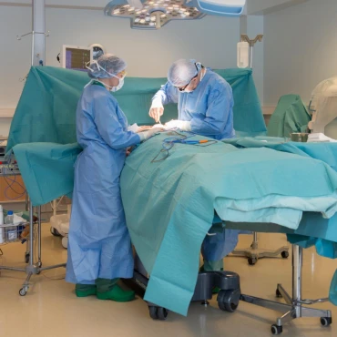
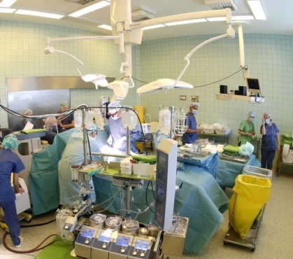
The surgical treatment of an aortic aneurysm consists of excision of the aneurysm, suturing of the defect, or its replacement with a vascular prosthesis. Taking into account the anatomical localization, aneurysms of the abdominal aorta, thoracic aorta, aortic arch, thoracoabdominal part of the aorta, and infrarenal aorta are resected.
In hemodynamically significant aortic insufficiency, resection of the ascending thoracic aorta is combined with aortic valve prosthesis. Endovascular prosthesis of aortic aneurysm with stent placement is an alternative to open vascular intervention.
Prognosis and prevention
The prognosis of aortic aneurysm is mainly determined by its size and concomitant atherosclerotic lesion of the cardiovascular system. In general, the natural course of aneurysms is unfavorable and is associated with a high risk of death from aortic rupture or thromboembolic complications. Aortic aneurysms with a diameter of 6 cm or more have a 50% annual probability of rupture, and smaller diameters have a 20% annual probability of rupture. Early detection and planned surgical treatment of aortic aneurysms is justified by low intraoperative (5%) mortality and good long-term results.
Preventive recommendations include controlling BP, organizing a proper lifestyle, regular follow-ups with a cardiologist and cardiac surgeon, and drug therapy for concomitant pathology. People at risk of developing aortic aneurysms should undergo a screening ultrasound examination.

