Coarctation of the aorta
Coarctation of the aorta has several types as well as different treatments. In this article, we will talk about what this disease is, in whom it is more common, what complications and features it has. We will also briefly analyze the diagnosis and therapy of this pathology.
The aorta is the largest vessel in the human body. It has several parts and departs from the left ventricle of the heart. The aorta forms many branches that supply blood to all tissues and organs of the human body.
So, coarctation (narrowing) of the aorta is a congenital malformation of the cardiovascular system. With this pathology, the aorta narrows, which entails insufficient blood supply to the lower body, including the kidneys. The latter secrete hormones that increase blood pressure. As a result, the vessels of the upper body and heart are subjected to even more stress.
The defect is usually located below the exit of the left subclavian artery from the aortic arch and above the junction of the descending aorta with the ductus arteriosus or arterial ligament.
Who has this disease most often?
Coarctation of the aorta is more common in certain genetic diseases (for example, Turner syndrome, Williams syndrome), as well as in newborns whose mothers have diabetes or take hydantoin-based drugs (these drugs can be used for epilepsy and other diseases of the nervous system)... Also, coarctation of the aorta occurs twice as often in males.
Classification
The disease is classified depending on the localization of the narrowing relative to the arterial ligament and there are three types of coarctation of the aorta:
- Preductal;
- Ductal;
- Postductal.
In these types, the narrowing is located above the arterial ligament, at its level and below it, respectively.
An isolated form of the disease, a combination of coarctation of the aorta with a defect of the interventricular septum, and a combination of the disease with complex congenital heart defects are also distinguished.
Less commonly used is the Bonnet classification. In accordance with it, the disease is divided into child and adult types, and the moment of appearance of symptoms that are significant for maintaining health is taken into account.
Could the disease be associated with another pathology?
Yes, the accompanying violations can be:
- aortic valve with two cusps instead of three;
- underdevelopment (hypoplasia) of the aortic arch;
- abnormal withdrawal of the right subclavian artery;
- intracerebral aneurysms;
- stenosis (narrowing) of the mitral valve, as well as pathology of other valves;
- Sean's syndrome (congenital defect of the left heart and aorta).
With the disease, collateral circulation develops bypassing stenosis (mainly from the subclavian, internal thoracic and intercostal arteries), an increase in blood pressure in the left ventricle, as well as in the vessels that are located above the site of narrowing. Over time, the patient develops an enlargement of the left ventricle (hypertrophy), heart failure and atherosclerosis of the coronary arteries.
Coarctation of the aorta causes an increase in blood pressure, and hypertension is one of the main risk factors for atherosclerosis. Therefore, with this disease, atherosclerotic lesions are often observed at an early age.
Symptoms of the disease
The clinical picture of coarctation of the aorta depends on the age of the patient, the location and degree of narrowing of the lumen, the presence and type of concomitant anomalies, as well as the rate of closure of the ductus arteriosus.
In severe preductal or ductal constriction, blood flow through the patent ductus arteriosus maintains blood supply to the lower body. At the same time, there is no difference in pressure and pulse between the upper and lower limbs (but the saturation on the right upper limb is higher than on the lower limb). After the closure of the ductus arteriosus, ischemia (a sharp lack of oxygen) of the abdominal organs occurs and heart failure develops, the heart rate in the lower extremities weakens or disappears.
Symptoms of the disease can be:
- decreased appetite;
- shortness of breath;
- tachycardia;
- hepatomegaly (enlarged liver);
- cyanosis of the skin of the lower body;
- increased sweating;
- decrease or absence of urine output;
- a change in the gas composition of blood and a change in its acidity.
Much less common is postductal aneurysm, in which blood flow to the lower body is disrupted immediately after birth. In a patient with this type of disease, symptoms of heart failure occur quickly and are severe.
In older children and adults, the disease is usually diagnosed when the causes of hypertension are identified. In this case, the following symptoms may occur:
- headache;
- dizziness, tinnitus, nosebleeds;
- intolerance to physical activity;
- shortness of breath;
- abdominal pain.
Diagnosis of the disease
Identification of pathology begins with examining the patient and interviewing him (if possible). During the examination, the doctor may find:
- absence of pulse or its weakening on palpation of the femoral artery;
- the difference in blood pressure between the upper and lower extremities;
- strengthening of the second tone at the point of aortic valve auscultation;
- systolic murmur over the base of the heart or continuous murmur in the interscapular region (a sign of the development of collateral circulation);
- trembling in the sternum;
- displacement of the apical impulse to the left and down (a sign of left ventricular hypertrophy).
The following diagnostic methods are also used:
- Pulse oximetry. With coarctation of the aorta, oxygen saturation measured in the lower limb will be lower than that measured in the upper limb.
- Measurement of blood pressure. This pathology manifests itself in different blood pressure on the arms and legs: the blood pressure on the upper limb will be higher than on the lower one.
- ECG. Signs of hypertrophy and overload of the ventricles of the heart can be found.
- X-ray of the chest. When carrying out this diagnostic method, it is possible to identify radiological signs characteristic of coarctation of the aorta, for example, an increase in the size of the heart and expansion of the ascending part of the aorta.
- Ultrasound. Allows you to visualize the location, structure and degree of narrowing, as well as accompanying violations.
- Angio-CT and angio-MRI. They are the most accurate methods for assessing the degree of coarctation, and are also used to determine the effectiveness of treatment and identify complications of surgical interventions.
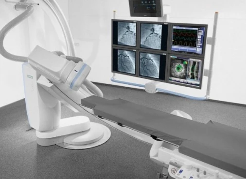
- Cardiac catheterization with aortography. Usually performed before surgery. Allows to measure the pressure gradient at the coarctation level.
Treatment
In newborns with preductal stenosis, surgery is performed immediately. For other types of the disease, planned treatment is possible.
Surgical treatment may include:
- removal of the narrowed area and connection of parts of the aorta end-to-end (Crawford's method);
- expansion of stenosis using a section extracted from the left subclavian artery (Waldhausen operation);
- operations using artificial materials and balloon plastic.
In adults, surgical treatment is indicated in the case of arterial hypertension, accompanied by significant stenosis (with a pressure difference between the right upper and lower extremities of more than 20 mm Hg). In this case, the disease should be confirmed by invasive examination (for example, cardiac catheterization with aortography). Basically, percutaneous procedures are performed (stenting, balloon angioplasty).
Disease prognosis
The pathology has a poor prognosis in newborns and infants with symptoms of heart failure, as well as in young patients who have not been treated. Lack of diagnosis at an early age leads to the persistence of arterial hypertension and the development of complications. Operated and non-operated patients are at increased risk of death from cardiovascular causes. The latter can be:
- heart failure;
- myocardial infarction;
- infections of the mediastinal organs;
- stroke;
- rupture, dissection, or infection of the aortic wall.
Summary
Coarctation of the aorta is a congenital heart disease and is of three types: preductal, ductal, and postductal. Most often, the disease occurs together with another pathology - a bicuspid aortic valve.
The clinical picture of coarctation of the aorta depends on several factors: the age of the patient, the location and degree of stenosis, concomitant anomalies, and the rate of closure of the ductus arteriosus. Common symptoms include high blood pressure in the upper body and a lack of pulse in the femoral arteries. In older children and adults, the disease is usually diagnosed when the causes of high blood pressure or headaches are identified.
The leading method of treatment is surgery. The preferred method for infants and young children is the Crawford method. In adults, surgical treatment is indicated in the case of arterial hypertension accompanied by significant stenosis.
With timely access to a doctor and proper treatment for coarctation of the aorta, the life expectancy of patients may not differ in any way from that of healthy people.
References:
- Harrison`s Principles of Internal Medicine 19/E (Vol.1). Dennis Kasper, Anthony Fauci, Stephen Hauseret all. McGraw-HillEducation 2015 ISBN: 0071802134 ISBN-13(EAN): 9780071802130.
- Interna szczeklika - duży podręcznik. Medycyna praktyczna. 2021. ISBN 9788374306522.
- Pediatria do LEK i PES. Anna Dobrzańska, Józef Ryżko. 2018. ISBN: 978-83-7609-855-5.


