Definition
An atrial septal defect is a congenital heart anomaly characterized by open communication between the right and left atria. An atrial septal defect is manifested by dyspnea, increased fatigue, physical retardation, palpitations, pale skin, heart murmurs, and frequent respiratory illnesses. An atrial septal defect is diagnosed, taking into account ECG data, echocardiography, catheterization of heart cavities, and angiopulmonography. In an atrial septal defect, the defect is sutured or plated, or an X-ray endovascular occlusion is performed.
General information
Atrial septal defect (ASD) is one or several holes in the septum separating the left and right atrial cavities, the presence of which causes pathologic discharge of blood and disturbance of intracardiac and systemic hemodynamics. In cardiology, atrial septal defect occurs in 5-15% of people with congenital heart disease, and it is diagnosed twice as often in women.
Along with ventricular septal defects, coarctation of the aorta, and open ductus arteriosus, atrial septal defects are among the most common congenital heart defects. It can be an isolated congenital heart defect or combined with other intracardiac anomalies: ventricular septal defect, abnormal pulmonary vein drainage, and mitral or tricuspid insufficiency.
Causes of atrial septal defect
The formation of the defect is associated with the underdevelopment of the primary or secondary interatrial septum and endocardial valleys during the embryonic period. Genetic, physical, environmental, and infectious factors can disrupt organogenesis.
The risk of developing an atrial septal defect in a future child is significantly higher in families with relatives with congenital heart disease. Cases of familial atrial septal defects in combination with atrioventricular block or underdevelopment of the hand bones (Holt-Oram syndrome) have been described.
In addition to hereditary causation, viral diseases of the pregnant woman (rubella, varicella, herpes, syphilis, etc.), diabetes mellitus and other endocrinopathies, taking certain medications and alcohol during pregnancy, occupational hazards, ionizing radiation, gestational complications (toxicosis, threat of miscarriage, etc.) can lead to the occurrence of atrial septal defect.
Hemodynamics in atrial septal defect
Due to the pressure difference between the left and right atria, an atrial septal defect causes arteriovenous shunting of blood from left to right. The amount of blood discharge depends on the size of the interatrial communication, atrioventricular resistance ratio, plastic resistance, and ventricular filling volume of the heart.
Blood shunting left-right is accompanied by increased filling of the small circulation circle, increased volume load of the right atrium, and increased work of the right ventricle. Due to the discrepancy between the area of the pulmonary artery valve opening and the volume of ejection from the right ventricle, relative pulmonary artery stenosis develops.
Classification of atrial septal defects
Atrial septal defects vary in the foramen’s number, size, and location.
Taking into account the degree and nature of the primary and secondary atrial septa underdevelopment, primary and secondary defects, as well as the complete absence of the interatrial septum resulting in a common, single atrium (three-chambered heart) are distinguished, respectively.
Primary ASDs include underdevelopment of the primary atrial septum with preservation of the primary interatrial communication. In most cases, they are combined with splitting the bicuspid and tricuspid valve flaps and an open atrioventricular canal. The primary atrial septal defect is usually large (3-5 cm), localized in the lower part of the septum above the atrial-ventricular valves, and having no lower edge.
Secondary ASDs are caused by the underdevelopment of the secondary septum. They are usually small (1-2 cm) and located in the center of the atrial septum or in the region of the vena cava orifices. Secondary atrial septal defects often combine with abnormal pulmonary veins flowing into the right atrium. In this type of malformation, the atrial septum is preserved in its lower part.
There are combined atrial septal defects (primary and secondary atrial septal defects, ASD in combination with a venous sinus defect). The atrial septal defect can also be part of the structure of complex CHD (pentalogy of Fallot) or combined with severe cardiac defects such as Ebstein’s anomaly, hypoplasia of the heart chambers, and transposition of the main vessels.
One variant of interatrial communication is an open oval window due to the underdevelopment of the foramen ovale’sintrinsic valve or its defect. However, an open foramen ovale is not a true septal defect associated with tissue insufficiency, so this anomaly cannot be classified as an interatrial septal defect.
Symptoms of atrial septal defect
Interatrial septal defects can occur with prolonged hemodynamic compensation, and their clinic is characterized by significant diversity. The severity of symptoms is determined by the size and localization of the defect, the duration of CHD, and the development of secondary complications. In the first month of life, the only manifestation of atrial septal defect is usually transient cyanosis during crying and restlessness, which is usually associated with perinatal encephalopathy.
In medium and large atrial septal defects, the symptomatology manifests itself already in the first 3-4 months or by the end of the first year of life and is characterized by persistent pallor of the skin, tachycardia, moderate lag in physical development, insufficient weight gain. Children with atrial septal defects are characterized by frequent respiratory diseases – recurrent bronchitis, pneumonia, prolonged wet cough, persistent dyspnea, abundant wet wheezing, etc., due to hypervolemia of the small circulation circle. In children in the first decade of life, frequent dizziness, tendency to faint, rapid fatigue, and dyspnea with physical exertion are distinguished.
Small atrial septal defects (up to 10-15 mm) do not cause impaired physical development or characteristic complaints in children, so the first clinical signs of the malformation may develop only in the second or third decade of life. Pulmonary hypertension and heart failure in atrial septal defects develop around 20 years of age, when cyanosis, arrhythmias, and rarely hemoptysis occur.
Diagnosis of atrial septal defect
An objective examination of a patient with an atrial septal defect reveals pallor of the skin, moderate stunting, and weight loss. On auscultation, a moderately intense systolic murmur is heard to the left of the sternum in the II-III intercostal spaces. In contrast to an interventricular septal defect or pulmonary artery stenosis, this murmur is never coarse.
In secondary atrial septal defects, ECG changes reflect right heart overload. Incomplete right bundle branch blockade, AV blockade, and sinus node weakness syndrome may be registered. Chest radiography allows you to see the strengthening of the pulmonary pattern, swelling of the pulmonary artery trunk, and increased shadow of the heart due to hypertrophy of the right atrium and ventricle. Fluoroscopy reveals a specific sign of an atrial septal defect—increased pulsation of the lung roots.
Echocardiography – study with color Doppler mapping reveals left-right blood discharge and the presence of atrial septal defect, which allows for the determination of its size and localization. Probing of heart cavities reveals increased pressure and blood oxygen saturation in the right heart and pulmonary artery. In case of diagnostic difficulties, the examination is supplemented with ventriculography, angiopulmonography, and cardiac MRI.
The atrial septal defect should be differentiated from interventricular septal defect, open ductus arteriosus, mitral insufficiency, isolated pulmonary artery stenosis, Fallot’s triad, and anomalous pulmonary vein drainage.
Treatment of atrial septal defect
Atrial septal defects are treated surgically only. The indication for cardiac surgery is the detection of hemodynamically significant arteriovenous effusion. The optimal age for correcting the defect in children is 1 to 12 years. Surgical treatment is contraindicated in cases of high pulmonary hypertension with venoarterial shunting due to sclerotic changes in pulmonary vessels.
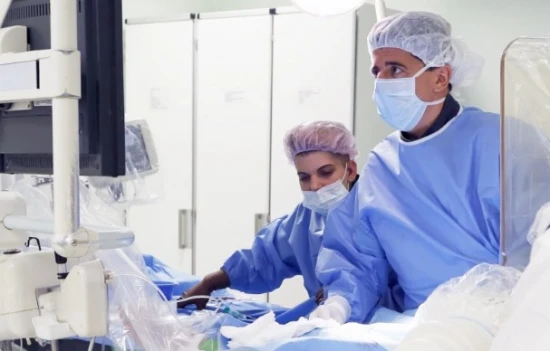
Various closure methods are used for atrial septal defects: suturing, repair with pericardial flap, or synthetic patch under hypothermia and IR. X-ray endovascular occlusion of the atrial septal defect allows the holes to be closed to not more than 20 mm.
Surgical correction of atrial septal defects is accompanied by good long-term results: 80-90% of patients have normalization of hemodynamics and absence of complaints.
Prognosis of atrial septal defect
Small atrial septal defects are compatible with life and can be detected even in old age. Some patients with a small atrial septal defect may have spontaneous closure of the orifice within the first five years of life. The life expectancy of individuals with large atrial septal defects in the natural course of the malformation is, on average, 35-40 years. Patients may die of right ventricular heart failure, cardiac rhythm and conduction disorders (paroxysmal tachycardia, atrial fibrillation, etc.), and, less frequently, of high-degree pulmonary hypertension.
Patients with atrial septal defects (operated and unoperated) should be monitored by a cardiologist and cardiac surgeon.


