Definition
Open atrioventricular canal defect is a complex intracardiac anomaly involving defects in the interventricular and interatrial septum and in both atrioventricular valves. The open atrioventricular canal leads to stunted physical development, dyspnea, tachycardia, fatigue, cyanosis, and frequent incidence of respiratory infections. Instrumental diagnosis of open atrioventricular canal includes ECG, echocardiography, chest radiography, cardiac cavity catheterization, and ventriculography. Surgical correction is indicated in the atrioventricular canal defect, i.e., ASD, VSD, mitral, and tricuspid valve repair.
General information
The open atrioventricular canal is a combined congenital heart defect characterized by the underdevelopment of intracardiac septa and atrial-ventricular valve anomalies. In cardiology, it accounts for 2-6% of all congenital heart defects. It is usually detected before the age of 1 year.
Open atrioventricular canal occurs in Down syndrome in 25-30% of cases. In 13-18% of patients, other cardiac anomalies are detected in addition to open atrioventricular canal: stenosis or atresia of the pulmonary artery, tetralogy of Fallot, coarctation of the aorta etc.
Causes of open atrioventricular canal
Embryologic abnormalities underlie the formation of the open atrioventricular canal. Factors that can cause underdevelopment of the septum and valves of the heart can be hereditary history of congenital malformations, chromosomal abnormalities, toxicosis of pregnancy, viral infections of the pregnant woman (rubella, herpes, chicken pox, influenza), endocrine disorders in the mother (diabetes mellitus), use of alcohol or drugs with teratogenic effect.
The structure of the defect in an open atrioventricular canal consists of the following components: atrial and ventricular septal defects and valve structure disorder (mitral and tricuspid).
Classification of open atrioventricular canal
There is a distinction between incomplete (partial), complete, and intermediate forms of open atrioventricular canal. Partially open atrioventricular canal accounts for 70% of malformation cases, while complete and intermediate forms account for 30%.
The incomplete form of the atrioventricular canal (primary atrial septal defect) is characterized by communication of the heart chambers at the level of the atria, as well as deformation or cleavage of the anterior mitral and/or septal tricuspid flaps.
The complete form of an open atrioventricular canal includes a merging defect of the membranous part of the atrial septum with a highly located defect of the atrial septum and the presence of one common valve between both halves of the heart. In intermediate forms of the malformation (oblique canals, Herbode’s defects), there are left ventricular-right atrial connections.
Hemodynamics in open atrioventricular canal
Intracardiac hemodynamic disorders in open atrioventricular canal depend on the anatomic form of the malformation. The partially open atrioventricular canal causes left-right shunting of blood; pulmonary hypertension is insignificant, and mitral insufficiency is moderately pronounced.
With a totally open atrioventricular canal, there is left-to-right shunt and a small right-to-left shunt of blood. Mitral and tricuspid insufficiency leads to systolic regurgitation of blood from the ventricles to the atria. Pulmonary hypertension and Eisenmenger’s syndrome develop early in this case. Intermediate forms of open atrioventricular canal are accompanied by blood flow from the left ventricle directly into the right atrium, right ventricular overload, and dilatation of the pulmonary trunk.
Symptoms of an open atrioventricular canal
Clinical manifestations of open atrioventricular canal develop in the first year of life, and subsequently, the condition of patients progressively worsens. There is a lag in the child’s physical development, rapid fatigue when feeding, decreased appetite, and pallor of the skin and mucous membranes. In the complete form of the open atrioventricular canal due to arterial hypoxemia, skin cyanosis is pronounced in the area of the nasolabial triangle, palms, and feet.
The presence of heart failure can be thought of as tachypnea, tachycardia, congestive wheezing in the lungs, and liver enlargement. Children with an open atrioventricular canal are prone to frequent colds, bronchitis, and pneumonia. By the age of 4-7 years, children develop an asymmetric chest deformity – a heart hump. Most children with a complete form of open atrioventricular canal die by the end of the 1st year of life.
Diagnosis of open atrioventricular canal
An open atrioventricular canal is usually diagnosed in the first days or months of a child’s life. Physical examination reveals enlarged heart borders, increased apex tremor, and systolic tremor above the apex. A characteristic auscultatory sign is hearing two noises different in localization and timbre: the systolic murmur of mitral valve insufficiency at the apex and the systolic murmur in the II-III intercostal spaces, as well as splitting of the II tone over the pulmonary artery.
To confirm the diagnosis of the open atrioventricular canal, ECG, cardiac ultrasound, cardiac cavity probing, chest radiography, and cardiac MRI are performed.
Electrocardiogram shows the leftward deviation of the heart’s electrical axis, left atrial hypertrophy, ventricular hypertrophy, right bundle branch block, or atrioventricular block. Radiography reveals cardiomegaly due to enlargement of both ventricles and the right atrium, increased pulmonary pattern, moderate bulging of the pulmonary artery arch, and signs of mitral insufficiency.
Echocardiography provides direct indications of an open atrioventricular canal: scanning reveals the presence of an ASD, VSD, clefting, anomalies of mitral valve leaflet attachment, and blood regurgitation in the valve area. The obtained data are confirmed by catheterization of heart cavities and ventriculography.
Comprehensive examination allows differentiating open atrioventricular canal from isolated atrial septal defect, interventricular septal defect, single ventricle of the heart, and transposition of the main vessels. It also excludes concomitant malformations—Fallot’s tetrad, open ductus arteriosus, and pulmonary artery stenosis—which is extremely important for proper treatment planning.
Treatment of open atrioventricular canal
Drug therapy for open atrioventricular canal is aimed at the prevention of infective endocarditis and the management of heart failure. Radical elimination of the malformation can only be performed surgically. Early surgical treatment is indicated for the complete form of open atrioventricular canal; in partial and intermediate forms, corrective surgery is performed at 6-10 years of age.
Correction of the incomplete form of the atrioventricular canal involves repairing the anterior mitral valve leaflet, closing the atrial septal defect, and, if necessary, repairing the tricuspid valve.
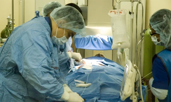
Correction of the complete open atrioventricular canal includes one-stage repair of the ASD, VSD, mitral, and tricuspid valves. In young children, palliative surgery for pulmonary trunk narrowing is sometimes performed beforehand. In intermediate variants of the open atrioventricular canal, VSD repair and tricuspid valve annuloplasty are performed. The operations are performed under cardiopulmonary bypass.
Prognosis in open atrioventricular canal
The course of the complete form of open atrioventricular canal is favorable: with timely correction of the malformation, 95% of patients die within the first five years of life. In the partial form of open atrioventricular canal, the average life expectancy of patients without surgery is 15-20 years. Intermediate forms are relatively favorable but also require surgical correction. Postoperative results of the open atrioventricular canal are satisfactory.
Failure to see a cardiologist or cardiac surgeon promptly increases the risk of cardiac complications – severe heart failure, arrhythmias, bacterial endocarditis, and high pulmonary hypertension, making surgical treatment impossible.

