Classification, pathophysiology of valvular pulmonary stenosis
Pulmonary stenosis is a defect in the valve of the pulmonary artery. In this case, the valve itself hardens, causing obstruction of the vessel and impaired blood flow. This disease is usually congenital, benign, diagnosed in children who have a positive prognosis. Pulmonary stenosis is also present in adults, usually in association with severe structural heart disease.
What is congenital pulmonary valve stenosis?
Pulmonary stenosis is a condition characterized by an obstruction in which blood flow from the right ventricle to the pulmonary trunk is interrupted. This obstruction is caused by a narrowing (called “stenosis” in medical parlance) at one or more points from the right ventricle to the pulmonary artery. Areas of potential stenosis include:
- thickening of the muscles under the valve of the pulmonary trunk;
- narrowing of the valve opening;
- or stenosis of the pulmonary trunk above the valve.
The most common form is obstruction of the valve itself, called pulmonary valve stenosis.
The normal pulmonary valve consists of three thin and flexible leaflets. When the right ventricle ejects blood, the normal valve leaflets open easily and do not obstruct the outflow of blood from the heart. Most often, with stenosis of the pulmonary valve, its leaflets thicken and merge together along the lines of separation (commissures). As a result, the leaflets become less pliable than normal, which contributes to the development of a narrowing of the hole and further disruption of blood flow.
When the pulmonic valve is blocked, the right ventricle must work harder to expel blood. To compensate for this extra load, the right ventricular muscle (myocardium) gradually thickens, a process called hypertrophy.
Reasons for the development of the disease
Pulmonary stenosis is commonly associated with congenital structural cardiac syndromes, including tetralogy of Fallot and Noonan's syndrome. Rubella virus infection during pregnancy is also a common cause of congenital stenosis, although it is not a genetic defect. Symptomatic onset of pulmonary stenosis in adults is also possible. It is sometimes seen during pregnancy if the stenosis is not previously identified, and also in women suffering from concomitant carcinoid syndrome. In addition, people with rheumatic heart disease, previous cardiothoracic surgery, or a heart tumor may develop pulmonary stenosis.
This defect accounts for 7-10% of all congenital heart anomalies. Pathology affects women to a slightly greater extent. About 2% of cases that occur in members of the same family do not end up with a genetic cause.
Pathophysiology
Isolated pulmonary stenosis is subdivided into valvular, subvalvular and supravalvular obstruction.
Valvular pulmonary stenosis is the most common type. In typical valvular disease, the adhesions are partially fused and the valve leaflets are thin. During systole, the right ventricle becomes domed. Less commonly, pulmonary valves may be dysplastic (underdeveloped), thickened, and non-fused. An associated hypoplastic annulus and upper pulmonary artery are common in atypical presentation and are also associated with Noonan syndrome.
Subvalvular pulmonary stenosis is a defect that obstructs the infundibular (subvalvular) region, i.e. significantly reducing its permeability. The cause of fibromuscular narrowing of the outflow tract of the right ventricle may be associated with a double-chambered right ventricle or Fallot's tetralogy, as well as surgical intervention on the valves.
Supravalvular pulmonary stenosis, also known as peripheral pulmonary stenosis, is essentially a functional obstruction of the pulmonary artery. Blockage is likely both in the basilar artery and at the bifurcation point, i.e. ramifications. It can also develop in the distal branches of the pulmonary artery or in any combination of areas. Supravalvular stenosis is associated with structural defects of the atrial or interventricular septum, patent ductus arteriosus, and tetralogy of Fallot. Supravalvular stenosis can also be the result of surgical correction of the transposition of the great arteries.
Signs and symptoms
Children with pulmonary stenosis usually do not have symptoms. They are outwardly healthy and do not show signs of any disease.
A heart murmur is the most common symptom found by a doctor, indicating a valve problem. Children with mild to moderate pulmonary valvular stenosis have an easily detectable heart murmur but are usually not clinically apparent.
In severe pulmonary valve obstruction, especially in critically ill neonates, the right ventricle cannot eject enough blood into the pulmonary trunk to maintain normal oxygen saturation. In these cases, venous, oxygen-poor blood bypasses the right ventricle. It flows from the right atrium to the left through the foramen ovale, or foramen, which is usually present in newborns. Thus, newborns with critical pulmonary stenosis will present with cyanosis (i.e., blue discoloration of the lips and nail beds) due to lower blood oxygen levels.
A newborn with critical pulmonary stenosis requires immediate treatment: either balloon dilatation (widening of the valve opening with the help of certain manipulations) or surgery.
In older children, a severe form of the disease can be manifested by rapid fatigue. Sometimes there is shortness of breath, especially with intense physical exertion. It is important that even a severe degree of stenosis rarely leads to the development of right ventricular failure, and also almost never provokes sudden death.
Diagnosis of the pulmonary stenosis
The first sign that suggests pulmonary stenosis is a heart murmur. An extraneous sound is a turbulent noise caused by the ejection of blood through an obstructive valve. Often there is a corresponding click: it is caused by the thick valve snapping into the open position.
The electrocardiogram in people with mild pulmonary stenosis is usually normal. With moderate to severe narrowing, an ECG film can reveal an increase in the right ventricle and thickening of its muscles.
Echocardiography is the most important non-invasive test for detecting and evaluating pulmonary valve stenosis. An echocardiogram accurately documents the presence of obstruction at the valve level, and a Doppler study is used to assess the degree of obstruction.
An echocardiogram is also important to rule out other problems that may be associated with pulmonary stenosis, such as an atrial or ventricular septal defect.
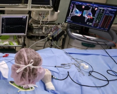
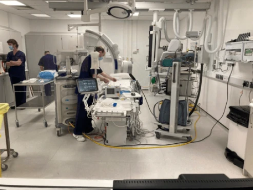
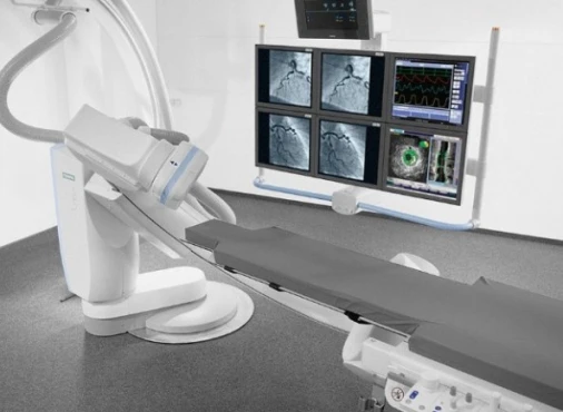
Cardiac catheterization is an invasive technique that allows doctors to accurately measure the degree of existing pulmonary stenosis. During cardiac catheterization, the pressure above and below the valve is measured to determine the degree of obstruction, and a video is taken to visualize the pulmonic valve. Angiography may be performed.
Over the past 15 years, echocardiography has largely replaced cardiac catheterization for diagnostic purposes. A complex invasive procedure is rarely required to make a diagnosis. Instead, balloon dilation is usually performed.
Treatment of valvular stenosis
Children with moderate pulmonary valve stenosis rarely need treatment. People with mild pulmonary valve stenosis are generally healthy. They can participate in all types of physical activity and sporting events and lead a normal life.
Mild pulmonary stenosis, diagnosed in childhood, rarely progresses beyond the first year of life, but still requires careful monitoring.
Children with moderate to severe pulmonary stenosis require treatment. Its type depends on the valve anomaly present. Most often, the pulmonary valve is of normal size, and the obstruction occurs due to fusion of commissures or lines of opening of the valve leaflets.
This "typical" form of pulmonary valve stenosis lends itself very well to balloon dilatation. This procedure is performed during cardiac catheterization and does not require open heart surgery. As a rule, in older children, the procedure is performed on an outpatient basis, unless otherwise indicated.
In neonates, balloon dilatation in critical pulmonary valve stenosis can be technically challenging. Often such children are born in a critical condition or easily pass into it.
Open-heart surgery is needed in difficult cases where they are occluded by thick and dysplastic leaflet tissue (eg, in patients with Noonan's syndrome).
In these cases, open-heart surgery may require valvotomy (opening the valve) or partial removal of its leaflet.
Disease prognosis
For children and adolescents with "typical" pulmonary stenosis, a balloon dilatation procedure is usually the only intervention needed. Rarely, older children experience recurrence of significant obstruction of the pulmonary valve after a successful procedure.
Neonates and young children with very severe valve obstruction also respond well to balloon dilatation, even if the valve is underdeveloped.
However, recurrence of significant pulmonary stenosis occurs in approximately 5-10% of children within 10 years of intervention. Sometimes these people may need a second balloon dilation or open-heart surgery.
It is important that all children with pulmonary stenosis, even after successful balloon dilatation or open-heart surgery, be evaluated regularly.
The prognosis for most patients is excellent. With the exception of critical stenosis in the neonatal period, most patients will lead a normal life. In 67% of patients undergoing revision surgery, 90% of maximal exercise tolerance is achieved, and only 15-20% require reoperation. 67% of reoperations are associated with significant pulmonary regurgitation - the reverse flow of blood from the artery.
Life of people with pulmonary valve stenosis
There are several options for how the disease can develop. The mildest is the one in which mild pulmonary stenosis, identified in childhood, remains the same in an adult, without becoming heavier. This means no progression and therefore no need for frequent medical interventions.
Most patients who have undergone balloon dilatation, and even open-heart surgery, do not need further attention from the doctor later.
With moderate stenosis, a person's well-being can also be quite good. A proportion of these people will develop complications and expert supervision will be recommended. Finally, pulmonary stenosis may present as part of a more complex set of congenital heart anomalies. All such patients require lifelong expert supervision and treatment.
References:
- Anderson K, Cnota J, James J, Miller EM, Parrott A, Pilipenko V, Weaver KN, Shikany A. Prevalence of Noonan spectrum disorders in a pediatric population with valvar pulmonary stenosis. Congenit Heart Dis. 2019 Mar;14(2):264-273.
- Khanra D, Shrivastava Y, Duggal B, Soni S. Congenital supravalvular and subvalvular pulmonary stenosis with hypoplastic pulmonary annulus associated with congenital rubella syndrome. BMJ Case Rep. 2019 Jul 10;12(7).
- Fathallah M, Krasuski RA. Pulmonic Valve Disease: Review of Pathology and Current Treatment Options. Curr Cardiol Rep. 2017 Sep 16;19(11):108.


